
9.2: Synaptic Control of Movement- Neuromuscular Junction and Motor Units
- Page ID
- 116200
This page is a draft and under active development. Please forward any questions, comments, and/or feedback to the ASCCC OERI (oeri@asccc.org).
- Describe the structural and functional aspects of motor neurons and the neuromuscular junction
- Explain the key steps of electrical excitation of muscle fibers
- Apply knowledge of motor units and motor unit recruitment to functional changes in muscle strength
Motor Neurons
Although the neuroanatomy of the motor system will be covered in a later section, it is important to realize that a large portion of the Central and Peripheral Nervous Systems contain collections of neurons that influence motor action in some way. Many of these neurons in the CNS belong to so-called, "motor circuits" and are classified as motor neurons as opposed to sensory or interneurons. Motor neurons located in cortical and subcortical regions of the forebrain, such as motor and premotor cortex and the basal ganglia are referred to as Upper Motor Neurons, while those located in the medulla and spinal cord are the Lower Motor Neurons. The function of the Upper Motor Neurons will be addressed in later sections. The Lower Motor Neurons of the spinal cord play a direct role in both triggering voluntary movement and maintaining muscle tone. These are the Alpha and Gamma motor neurons, respectively.
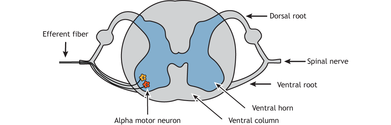
The cell bodies of the Alpha motor neurons are located in the central nervous system in the ventral (i.e. anterior) horn of the spinal cord. Their axons leave the spinal cord via the ventral roots and travel primarily to skeletal muscle via efferent (outgoing) spinal nerves to cause muscle contraction. Gamma motor neurons function to keep muscle fibers prepared for action by stimulating them in a way to maintain "tautness" or muscle tone. This resting muscle tonus allows the muscle to respond much more effectively to the alpha motor neuron stimulation, thus alpha and gamma neurons work together to maintain a muscle's sensitivity for movement. Just imagine how difficult it would be to shoot a rubber band across a room if it wasn't stretched or taut (Gamma motor neuron influence) prior to letting it go (Alpha motor neuron influence).
The Neuromuscular Junction
A unique specialization of the skeletal muscle is the site where a motor neuron’s axon terminal meets the muscle fiber—called the neuromuscular junction (NMJ). This is where the muscle fiber first responds to signaling by the motor neuron. Every skeletal muscle fiber in every skeletal muscle is innervated by a motor neuron at the NMJ. Excitation signals from the neuron are the only way to functionally activate the fiber to contract. It is important to note that one motor neuron can activate multiple muscle fibers due to axonal branching (see the Motor Units section below for more details on this topic).
Excitability of Muscle: Excitation-Contraction Coupling
All living cells have membrane potentials, or electrical gradients across their membranes. For example, in neurons which are not being stimulated the membrane potential is approximately -70 mV inside of the cell relative to the outside. This is referred to as a neuron’s resting potential.
Although living cells have a cellular membrane, only some, such as neurons and muscle cells are excitable. In other words, they can shift quickly from a resting state to an excited one. Thus, neurons and muscle cells can use their membrane potentials to generate electrical signals. They do this by controlling the movement of charged particles, called ions, across their membranes to create electrical currents. This is achieved by opening and closing specialized proteins in the membrane called ion channels. Although the currents generated by ions moving through these channel proteins are very small, they form the basis of both neural signaling and muscle contraction.
Both neurons and skeletal muscle cells are electrically excitable, meaning that they are able to generate action potentials. An action potential is a special type of electrical signal that can travel along a cell membrane as a wave. This allows a signal to be transmitted quickly and faithfully over long distances.
Although the term excitation-contraction coupling confuses or scares some students, it comes down to this: for a skeletal muscle fiber to contract, its membrane must first be “excited”—in other words, it must be stimulated to fire an action potential. The muscle fiber action potential, which sweeps along the muscle fiber as a wave, is “coupled” to the actual contraction through the release of calcium ions (Ca++) from the muscle cells. Once released, the Ca++ interacts with the shielding proteins, forcing them to move aside so that the actin-binding sites are available for attachment by myosin heads. The myosin then pulls the actin filaments toward the center, shortening the muscle fiber.
In skeletal muscle, this sequence begins with signals from the somatic motor division of the nervous system. In other words, the “excitation” step in skeletal muscles is always triggered by signaling from the nervous system.
.jpg?revision=1)
Although a small number of motor neurons activating the skeletal muscles of the face, head, and neck are located in the brainstem, most motor neurons originate in the spinal cord, directing skeletal muscle fibers to contract throughout the rest of the body. These neurons have long processes, called axons, which are specialized to transmit action potentials long distances— in this case, all the way from the spinal cord to the muscle itself (which may be up to three feet away). The axons of multiple neurons bundle together to form nerves, like wires bundled together in a cable.
Signaling begins when a neuronal action potential travels along the axon of a motor neuron, and then along the individual branches to terminate at the NMJ. At the NMJ, the axon terminal releases a chemical messenger, or neurotransmitter, called acetylcholine (ACh). The ACh molecules diffuse across a minute space called the synaptic cleft and bind to ACh receptors located within the motor end-plate of the sarcolemma on the other side of the synapse. Once ACh binds, a channel in the ACh receptor opens and positively charged ions can pass through into the muscle fiber, causing it to depolarize, meaning that the membrane potential of the muscle fiber becomes less negative (closer to zero).
As the membrane depolarizes, another set of ion channels called voltage-gated sodium channels are triggered to open. Sodium ions enter the muscle fiber, and an action potential rapidly spreads (or “fires”) along the entire membrane to initiate excitation-contraction coupling.
Things happen very quickly in the world of excitable membranes (just think about how quickly you can snap your fingers as soon as you decide to do it). Immediately following depolarization of the membrane, it repolarizes, re-establishing the negative membrane potential. Meanwhile, the ACh in the synaptic cleft is degraded by the enzyme acetylcholinesterase (AChE) so that the ACh cannot rebind to a receptor and reopen its channel, which would cause unwanted extended muscle excitation and contraction.
Propagation of an action potential along the sarcolemma is the excitation portion of excitation-contraction coupling.This excitation actually triggers the release of Calcium ions (Ca++) from their storage in the cell’s sacroplasmic reticulum (SR). For the action potential to reach the membrane of the SR, there are periodic invaginations in the sarcolemma, called T-tubules (“T” stands for “transverse”). The diameter of a muscle fiber can be up to 100 μm, so these T-tubules ensure that the membrane can get close to the SR in the sarcoplasm. The arrangement of a T-tubule with the membranes of SR on either side is called a triad. The triad surrounds the cylindrical structure called a myofibril, which contains actin and myosin.
The T-tubules carry the action potential into the interior of the cell, which triggers the opening of calcium channels in the membrane of the adjacent SR, causing Ca++ to diffuse out of the SR and into the sarcoplasm. It is the arrival of Ca++ in the sarcoplasm that initiates contraction of the muscle fiber by its contractile units, or sarcomeres.
Motor Units
As described in the Neuromuscular Junction section above, every skeletal muscle fiber is innervated by an axon terminal of a motor neuron in order to contract. Each muscle fiber is innervated by only one motor neuron, but each motor neuron innervates a group of muscle fibers due to the axon's ability to branch. Recall that the NMJ is the point of connection between each axon terminal and it's corresponding muscle fiber. A motor neuron and the group of muscle fibers in a muscle that it innervates is called a motor unit. The size of a motor unit is variable depending on the nature of the muscle.
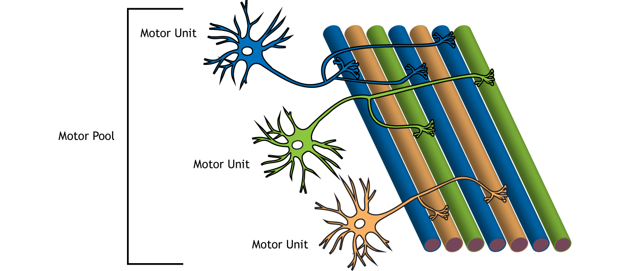
A small motor unit is an arrangement where a single motor neuron supplies a small number of muscle fibers in a muscle. Small motor units permit very fine motor control of the muscle. The best example in humans is the small motor units of the extraocular eye muscles that move the eyeballs. There are thousands of muscle fibers in each muscle, but every six or so fibers are supplied by a single motor neuron, as the axons branch to form synaptic connections at their individual NMJs. This allows for exquisite control of eye movements so that both eyes can quickly focus on the same object. Small motor units are also involved in the many fine movements of the fingers and thumb of the hand for grasping, texting, etc.
A large motor unit is an arrangement where a single motor neuron supplies a large number of muscle fibers in a muscle. Large motor units are concerned with simple, or “gross,” movements, such as powerfully extending the knee joint. The best example is the large motor units of the thigh muscles or back muscles, where a single motor neuron will supply thousands of muscle fibers in a muscle, as its axon splits into thousands of branches.
There is a wide range of motor units within many skeletal muscles, which gives the nervous system a wide range of control over the muscle. The small motor units in the muscle will have smaller, lower-threshold motor neurons that are more excitable, firing first to their skeletal muscle fibers, which also tend to be the smallest. Activation of these smaller motor units results in a relatively small degree of contractile strength (tension) generated in the muscle. As more strength is needed, larger motor units, with bigger, higher-threshold motor neurons, are enlisted to activate larger muscle fibers. This increasing activation of motor units produces an increase in muscle contraction known as recruitment.
As more motor units are recruited, muscle contraction grows progressively stronger. In some muscles, the largest motor units may generate a contractile force of 50 times more than the smallest motor units in the muscle. This allows a feather to be picked up using the biceps brachii arm muscle with minimal force, and a heavy weight to be lifted by the same muscle by recruiting the largest motor units.
When necessary, the maximal number of motor units in a muscle can be recruited simultaneously, producing the maximum force of contraction for that muscle, but this cannot last for very long because of the energy requirements to sustain the contraction. To prevent complete muscle fatigue, motor units are generally not all simultaneously active, but instead some motor units rest while others are active, which allows for longer muscle contractions. The nervous system uses recruitment as a mechanism to efficiently utilize a skeletal muscle.
Attributions:
- Sections on Motor Neurons, Neuromuscular Junction, and Motor Units adapted from J. Gordon Betts, Kelly A. Young, James A. Wise, Eddie Johnson, Brandon Poe, Dean H. Kruse, Oksana Korol, Jody E. Johnson, Mark Womble, Peter DeSaix, Anatomy and Physiology, OpenStax. License: CC BY 4.0
- ‘Alpha Motor Neurons’ and 'Motor Unit and Pool‘ graphics by Casey Henley. License: CC BY-NC-SA 4.0 International License.