
1.2: History of Biopsychology
- Page ID
- 120903
This page is a draft and under active development. Please forward any questions, comments, and/or feedback to the ASCCC OERI (oeri@asccc.org).
- Discuss the history of biopsychology.
- Discuss the important contributors to biopsychology and the contributions each made.
- Explain the current direction of biopsychology.
Overview
Biopsychology as a scientific discipline is relatively new. However, early philosophers such as Hippocrates wrote about the importance of biological factors in human behavior. In this section, we will examine the emergence of biopsychology as a scientific field. We will further outline important events and contributions to biopsychology, beginning with early philosophers and their understanding of the biological aspects of behavior and mental processes. We then examine a number of more recent historical figures who made major contributions to the development of biological psychology and discuss their contributions to the discipline. Lastly, we will look at current directions in the field.
Biopsychology as a Discipline
Biopsychology as a scientific discipline emerged from a variety of scientific and philosophical traditions in the 18th and 19th centuries. Although the exact date of inception of biological psychology is unknown, there have been a number of milestones in its emergence. William James in his book, The Principles of Psychology (1890), argued that the scientific study of psychology should be grounded in an understanding of biology (Walinga, 2014). Like many early psychologists, James had extensive training in physiology.
In An Outline of Psychobiology, Knight Dunlap used the term "psychobiology" to explain the role of biology in behavior. In 1949, Donald Hebb wrote his influential book, The Organization of Behavior, where he introduced the first comprehensive theory on how the brain might create and control complex psychological functioning, such as thought, memories, emotions and perceptions. In other words, even before biopsychology was formally recognized as a branch of psychology or as a discipline, the connection between psychology and biology was recognized. With case studies such as Phineas Gage and invention of methods such as the electroencephalogram (EEG) and computed tomography (CT or CAT) scans, scientists began to link the brain to specific behavior and cognition as biopsychology as a discipline began to emerge.
The Origins of Biopsychology
It is important to examine the historical path of our understanding of the brain and its role in our behavior and mental processes. Examining the history of biopsychology allows us to understand its development over time, highlighting instances where researchers were wrong about the nature of brain-behavior relationships and revealing what we have yet to explain (Saucier and Elias, 2006). Studying the history of a scientific discipline gives us a roadmap of where we have traveled from and in what direction we need to go.
Early Views of Brain and Behavior
Early philosophers, such as Aristotle (384-322 B.C.E.), believed that one's mind resided in the heart. He believed that since our blood started from the heart, the soul also originated there. Plato (428-347 B.C.E.) argued that the executor of reason was the heart and our animalistic desires and emotions were controlled by the liver (Gross, 1987). Many ancient cultures, including the Chinese, Indian, and Egyptian also shared the same belief (Carlson, 2014). When the Egyptians embalmed a person, the heart was saved and buried with the individual but the brain was discarded (Klein and Thorne, 2006). However, there were early Greeks, such as Hippocrates (460-377 B.C.E.) , who believed that it was the brain and not the heart where the locus of the mind resided. He wrote: "It ought to be generally known that the source of our pleasure, merriment, laughter, and amusement, as of our grief, pain, anxiety, and tears is none other than the brain. It is specially the organ which enables us to think, see, and hear......It is the brain too which is the seat of madness and delirium, of the fears and frights which assail us" (Gross, 1987, p. 843-844).
During the 3rd Century B.C., in Alexandria, a science museum was established. Since human dissection was practiced in that city for centuries, the anatomical study of the human body flourished (Gross, 1987). Two neuroanatomists, Herophilos and Erasistratos, contributed to our knowledge of the human brain. Herophilos distinguished the cerebellum (at the very base of the back of the brain) and the cerebrum (the two cerebral hemispheres). He hypothesized that since the cerebellum was denser than the other parts of the brain, it must control the muscles (a guess of impressive accuracy). And he provided the first clear description of the cavities within the brain known as ventricles (Figure 1.2.1). Erasistratos continued the work of Herphilos and proposed that human intelligence was related to the number of convolutions (ridges) in the brain; the more convolutions an individual's brain had, the more intelligent that person would be.
During the Roman Empire, the Greek anatomist Galen (A.D. 130-200) dissected the brains of sheep, monkeys, dogs, pigs, among other non-human mammals (Carlson, 2014). He believed the brain to be the site of sensation and thought, and the controller of movement (Gross, 1987). He stated that spinal cord was an extension of the brain and chronicled the relationship between the spinal nerves and specific muscles each controlled. For the next advance in understanding spinal function, we must await Bell and Magendie in the 19th Century.
Middle Ages

Avicenna (980-1037 C.E.) was a Persian physician who some regard as the father of modern medicine. He recognized biological psychology in his treatment of illnesses that involved emotions (Syed, 2002). In his book, The Canon of Medicine (Qanun: Law of Medicine), Avicenna dedicated three chapters to neuropsychiatric disorders where he defined madness (Junun) as originating in the middle part of the brain. He discussed a condition similar to schizophrenia, which he called Junun Mufrit, characterized by agitation, behavioral and sleep disturbances, giving inappropriate answers to questions, and an occasional inability to speak. Avicenna also discovered the cerebellar vermis, which he simply called the vermis, and the caudate nucleus. Both terms are still used in neuroanatomy today (Mohamed, 2008). He further explained that "humidity" inside the head contributed to mood changes and when this moisture inside the brain goes beyond limits, the brain loses control over its rationality and mental disorders appear (Haque, 2004). Lastly he presented detailed knowledge about skull fractures and their surgical treatments (Aciduman, Arda, Ozakturk, and Telatar, 2009). He also discussed phenomena such as insomnia, mania, hallucinations, nightmares, dementia, epilepsy, stroke, paralysis, vertigo, melancholia, and tremors (Mohamed, 2008). He was also the first person to associate mental deficits with deficits in the brain's middle ventricle or frontal lobe (Mohamed, 2008).
Renaissance
By the beginning of the 16th century, Andreas Vesalius, founder of modern anatomy, rekindled interest in neuroscience (Gross, 1987). Through his dissection of human cadavers, Vesalius found problems with the Galenic view of anatomy (which emphasized three major systems--the heart, brain, and blood--and the importance of a balance between four bodily fluids--blood, phlegm, and black and yellow bile). Vesalius published seven books, with the fourth and seventh books devoted to the nervous system (Van Laere, 1993). Vesalius outlined seven pairs of 'brain nerves', each with a specialized function. In his seventh book which was solely devoted to the brain, he successfully describes the cerebral membranes, the ventricular system, and the cerebrum (Van Laere, 1993). Other scholars furthered Vesalius' work by adding their own detailed sketches of the human brain.
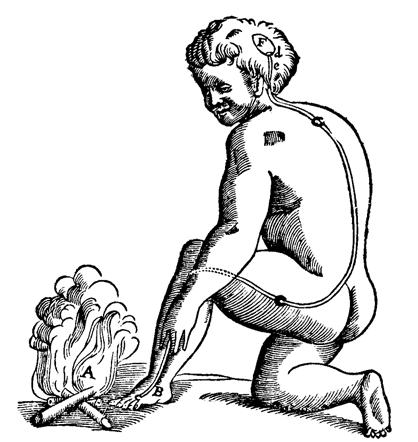
René Descartes (1596-1650) proposed the reflexive theory in terms of our behavior (Elias and Saucier, 2006). This theory explained reflexive behavior as an "external stimuli [that] would move the skin, in turn moving the filaments, releasing the animal spirits and innervating the muscles" (Elias and Saucier, 2006, p 6). His mechanical explanation of movement without conscious intervention allowed for scientific study of behavior. Furthermore, his theory encouraged using animal models to study biological bases of behavior (Cobb, 2002). He also studied the physiology of the brain, proposing the theory of dualism (the view that mind and brain are separate) to tackle the issue of the brain's relation to the mind. He suggested that the pineal gland was where the mind interacted with the body after recording the brain mechanisms responsible for circulating cerebrospinal fluid (Elias and Saucier, 2006).
Discoveries of the 19th Century
Localization of Function
Starting in early 1800's, a series of researchers began to catalog different brain parts and their presumed role in behavior. In 1811, Jean-Cesar Legallois (1770-1840) discovered that when he destroyed tissue in the medulla, a procedure called lesioning, there was an immediate cessation of respiration (Elias and Saucier, 2006). Charles Bell (1774-1842) and Francois Magendie (1783-1840) discovered the different function of spinal cord nerves where the ventral roots transmitted motor impulses and posterior roots received sensory input (Bell-Magendie Law). Given the specialization found in spinal cord nerves, Bell recommended that further investigation of the the entire nervous system should examine functional and anatomical segregation (Elias and Saucier, 2006).
Taking on the challenge, Franz Joseph Gall (1758-1828) and Johann Spurzheim (1776-1832) formulated phrenology where measurement of the skull was used to determine personality characteristics of an individual. Gall made the claim that here were twenty-seven distinct cognitive abilities that could be localized on the cortex of the human brain (the cortex composing the outermost part of the brain). He believed that the design of the brain changed as each of us developed certain characteristics and this resulted in corresponding changes in the skull. Where the skull rises, one can see excellence in the human qualities associated with the underlying brain and where there were depressions in the skull, human defects were represented (Lau, Loi and Abdullah, 2021). Measuring the skull by using a technique called cranioscopy would allow the scientist to detect deformation and bumps on the skull which would outline the person's personality (Elias and Saucier, 2006).
In 1848, John Martyn Harlow treated Phineas Gage and documented his case. Gage, who was a railroad worker, had his frontal lobe pierced by an iron tamping rod in a blasting accident. He survived the trauma but suffered extensive damage to his left prefrontal cortex (Macmillan, 2001). Through Gage's case study, Harlow showed the connection between the prefrontal cortex damage, executive functioning, and personality changes.
In 1861, Paul Broca (1824-1880) presented a case where a young man had sustained damage to the left frontal lobe and as a result was unable to produce spoken speech. However, the patient was able to comprehend speech and had average intelligence. After his death, Broca performed an autopsy and determined that the patient had a lesion in the frontal lobe in the left cerebral hemisphere. Broca collected similar cases of damage to the left frontal lobe with the patient losing their ability to speak (Lau, Loi and Abdullah, 2021). One such case was Phineas Gage who lost his speech after his accident. Broca published his findings from the autopsies of twelve patients in 1865. Today, we recognize Broca's Area in the frontal lobe as a critical region in speech production. His work inspired others to perform careful autopsies with the aim of linking more brain regions to sensory and motor functions.
Carl Wernicke (1848-1904) further developed the theory of the specialization of specific brain structures in language comprehension and production. He wrote a paper in 1874, suggesting that housed in the left temporal lobe was an auditory comprehension center. He stated that when this area, now known as Wernicke's area, was damaged, the individual could not comprehend other's speech or use words correctly (Elias and Saucier, 2006). The concept of "localization of function", specific functions residing in specific parts of the brain, was established the end of the 19th century. Respiration and the medulla and the roles of Broca's and Wernicke's areas in language are examples of this concept.
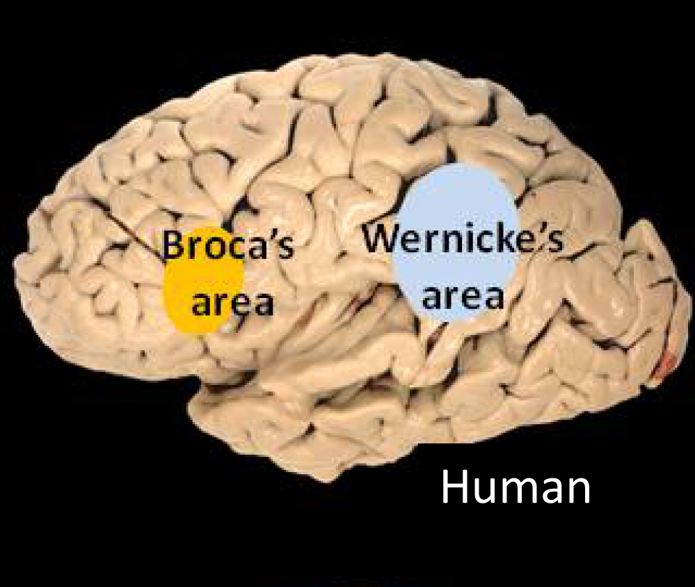 Figure \(\PageIndex{5}\): The two language areas of the brain. (Public Domain; via Wiki media Commons)
The Neuron
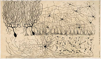
The last monumental neurological discovery of the 19th century was the identification of the specific structural unit responsible for these brain functions, the neuron. Studies of the brain became more sophisticated after the invention of the microscope and the development of a staining procedure by Camillo Golgi during late 1873 (Elias and Saucier, 2006). Golgi (1843-1926) was a histologist (one who studies the cellular structure of tissue) and a physician (Klein and Thorne, 2006). He used a silver chromate salt to reveal the intricate structures of single neurons (Elias and Saucier, 2006). Through this technique, he was able to reveal that the neuron has three distinct structures: dendrite, cell body, and axon (Elias and Saucier, 2006). Furthermore, he revealed that the axon can be short or very long, enabling the cell body to send messages to neurons far away. Therefore the axon must be the output carrier for the neuron. This technique is called the Golgi stain, named in his honor.
The Golgi stain technique was used by Santiago Ramón y Cajal to outline the neural connections in the brain (Elias and Saucier, 2006). While Golgi believed that neurons were fused together to form a continuous circuit, Cajal demonstrated that neurons are not physically connected but do have a mechanism for communicating (Elias and Saucier, 2006). This led to the formation of the neuron doctrine, the hypothesis that the functional unit of the brain is the neuron, also known as a nerve cell. Today, the field accepts neuronal theory which states that the nervous system is made up of individual nerve cells called neurons (Klein and Thorne, 2006).
Golgi and Ramón y Cajal shared the Nobel Prize in Physiology or Medicine in 1906 for their extensive observations, descriptions, and categorizations of neurons throughout the brain. Golgi was praised for his staining technique and Cajal for developing the neuron doctrine.
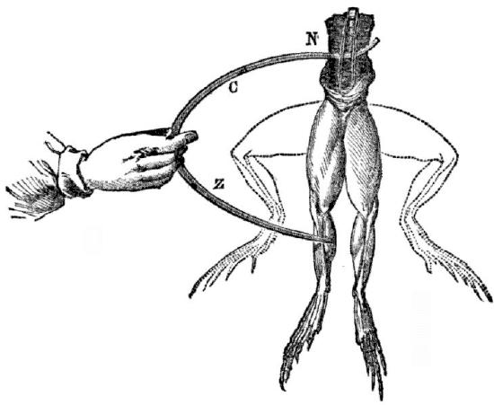
One of the most important questions given Cajal's neuron doctrine was the mechanism for communication between neurons. The role of electricity in nerves and electrical communication between neurons was first observed in dissected frogs by Luigi Galvani. In 1780, he showed that an electrical stimulus applied to the motor nerve of a frog's nerve cell produced a twitch. This experiment suggested that electricity was an important functional element of the nervous system (Klein and Thorne, 2006).
Discoveries of the 20th Century
The Synapse
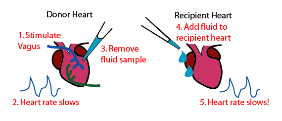
Charles Scott Sherrington (1857-1952) accepted Cajal's neuron doctrine and set out to discover the point of functional contact between neurons. He called this connection point the synapse, which means "binding together" (Klein and Thorne, 2006). His discovery was made by studying spinal reflexes in dogs. Confirming Galvani's electrical theory, he demonstrated the electrical transmission between neurons (Elias and Saucier, 2006). He received the Nobel Prize in 1932 for his research. A German pharmacologist and physician, Otto Loewi (1873-1961), investigated chemical communication between neurons. He removed the heart of two frogs and placed them in separate containers, each one filled with a chemical solution that allowed the hearts to continue to beat. He stimulated the vagus nerve of the first heart which slowed down its beating. He shared the solution from the first heart's container with the second heart and the beating of the second heart also slowed. He concluded that when he stimulated the vagus nerve of the first heart, a chemical was released to slow down its beating and when the solution from the first heart's container was shared with the second heart, the chemical directed the second heart to also decrease its beating (Klein and Thorne, 2006). He originally called this chemical vagusstoff, referring to its secretion after the stimulation of vagus nerve. This chemical was later identified as a neurotransmitter, acetylcholine (ACh). ACh was originally identified in 1915 by Henry Hallett Dale. However, Loewi is credited with establishing its role in chemical communication between nerve cells (Klein and Thorne, 2006).
Today, dozens of neurotransmitters have been identified in the brain. After acetylcholine, noradrenaline (now known as norepinephrine) was discovered in the 1940's, followed by dopamine and serotonin in the next decade and gamma-aminobutyric acid (GABA), glutamate, and glycine in the 1960's (Wickens, 2021). Later discoveries of neuropeptides, neuromodulators, and certain gases have added to the complexity of chemical communication in the brain. Our understanding of the impact and role of these chemical messengers and their effect on behavior and mental processes has given rise to the field of psychopharmacology.
Neural Conduction
Previous research had clearly demonstrated that electrical impulses played a role in neural communication. But the finer details of how this was occurring had not been discovered (Wickens, 2021). A series of discoveries led to our present understanding of neural conduction.
In 1902 and again in 1912, Julius Bernstein (1836-1917) was the first researcher to discover the action potential - the unique electrical signal traveling the length of an axon (Elias and Saucier, 2006). Additionally, he demonstrated that this electric conduction was due to change in ionic concentration of the cell's intracellular fluid. However, given the microscopic size of the cell, it was difficult to measure and study mammalian cells. In 1936, John Z. Young, an Oxford biologist, located a neuron in the body of a squid. The axon was nearly 1 mm in diameter which was 100 to 1,000 times larger than a mammalian axon and could be studied for about six hours after its dissection (Elias and Saucier, 2006). In 1952, two Cambridge University physiologists, Alan Hodgkin and Andrew Huxley published a set of papers describing their research on the giant squid axon. Much of our understanding of neural functioning came from the giant squid. Hodgkin and Huxley created a technique to measure electrical changes of the neuron by inserting an electrode inside and one positioned outside of the neuron without damaging it. This enabled them to measure changes in electrical charge when the neuron was at rest and when it produced an action potential (Wickens, 2021). A detailed discussion of both concepts, resting and action potential will follow in future chapters.
Studying Brain Activity and Brain Imaging
With the invention of machines such as the electroencephalograph (EEG), CT scan, and magnetic resonance imaging (MRI), scientists were able to peak into the activity and images of patients' brains while they were alive and engaged in mental activity. In 1924, Hans Berger recorded the first human brain EEG. This machine allowed researchers to study the overall electrical activity of the brain. In 1953, Kleitman and Aserinsky discovered Rapid Eye Movement (REM) using EEG on sleeping subjects. Today EEGs are helpful in diagnosing epilepsy and sleep disorders. Many credit their work as the beginning of modern study of sleep. The first image of the brain by CT scan was in 1971 by Godfrey Hounsfield. He used the CT scan to locate a brain tumor (Wells, 2005). CT scans can be used to locate brain tumors, injury, or atrophy. However, the CT scan uses Xrays to provide an image of the brain which could pose risk to brain tissue. Magnetic resonance imaging (MRI) was also used in 1970's to give pictures of the brain. An MRI does not use Xrays and is therefore found to be a safer method for producing human brain scans.
In 1950, positron emission tomography (PET) scans were used to look at brain activity or communication in a living patient. Researchers were able to look at different parts of the brain in different situations. In other words, they could look at real time activity of the brain as the person laughed or was scared to see what ares of the brain lit up. This gave an indication of what areas of the brain maybe responsible for different emotions or behaviors. The problem with PET scans was that an individual receiving a PET scan had to drink or be injected with a mildly radioactive substance, called a tracer. Once in the bloodstream, the amount of tracer in any given region of the brain can be monitored. As brain areas become more active, more blood flows to that area. A computer monitors the movement of the tracer and creates a rough map of active and inactive areas of the brain during a given behavior. PET scans show little detail, are unable to pinpoint events precisely in time, and require that the brain be exposed to radiation. In 1990s, a group in Bell Laboratories invented functional Magnetic Resonance Imaging (fMRI). This machine shows changes in brain activity over time by tracking blood flow and oxygen levels. The fMRI provides more detailed images of the brain’s structure, as well as better accuracy in time, than is possible in PET scans.
Today, these instruments have greatly contributed to our understanding of the brain and its function in behavior and cognition. With their high level of detail, MRI and fMRI are often used to compare the brains of healthy individuals to the brains of individuals diagnosed with psychological disorders. This comparison helps determine what structural and functional differences exist between these populations.
Today
In April 2013, the BRAIN (Brain Research through Advancing Innovative Neurotechnologies) Initiative was launched. The focus of this initiative was to advance our understanding of the human brain. In the fiscal year 2020, 10.1 billion U.S. dollars has been allocated to neuroscience studies (Mikulic, 2021). Different institutions, agencies and foundations such as Food and Drug Administration, Defense Advanced Research Projects Agency, have joined in this effort.
References
Aciduman, Ahmet, Arda, Berna, Ozakturk, Fatma G., Telatar, Umit F. (2009). What does Al-Qanun Fi Al-Tibb (the Canon of Medicine) say on head injuries?. Neurosurgical Review, 32 (3), 255-263.
Carlson, Neil R. (2014). Foundations of Behavioral Neuroscience, 9th Edition. Pearson Publishing.
Cobb, Matthew (2002). Timeline: Exorcizing the animal spirits: Jan Swammerdam on nerve function. Nature Reviews Neuroscience, 3 (5), 395-400.
Gross, Charles G. (1987). Neuroscience, Early History of. In Adelman, George (ed.), Encyclopedia of Neuroscience, Kirkhauswer Verlag AG, pp. 843-847.
Haque, Amber (2004). Psychology from Islamic perspective: Contributions of early Muslim scholars and challenges to contemporary Muslim psychologists. Journal of Religion and Health, 43 (4), 357-377.
Klein, Stephen B. and Thorne, B. Michael (2006). Biological Psychology. Worth Publishers.
Lau, Yoke Lian, Loi, Chek Kim and bin Abdullah, Mohd Nor Azan (2021). The Historical Development of the study of Broca's Aphasia. Malang Neurology Journal, 7 (2), 125-128.
Macmillan, Malcolm (2001). John Martyn Harlow: Obscure country physician? Journal of the History of Neurosciences: Basic and Clinical Perspectives, 10 (2), 149-162.
Mikulic, Matej (2021). Total neuroscience funding by the National Institutes for Health (NIH). Statica.com. https://www.statista.com/statistics/...isticContainer
Mohamed, Wael MY (2008). History of Neuroscience: Arab and Muslim contributions to modern neuroscience. IBRO History of Neuroscience: 255.
Syed, Ibrahim B. (2002). Islamic medicine: 1000 years ahead of its times. Journal of the International Society for the History of Islamic Medicine 2, 2-9.
Van Laere J. Vesalius en het zenuwstelsel [Vesalius and the nervous system]. Verh K Acad Geneeskd Belg. 1993;55(6):533-76. Dutch. PMID: 8209578.
Wells, P. N. T. (2005). “Sir Godfrey Newbold Hounsfield KT CBE. 28 August 1919 – 12 August 2004: Elected F.R.S. 1975”. Biographical Memoirs of Fellows of the Royal Society. 51: 221–235. doi:10.1098/rsbm.2005.0014.
Wickens, A. P. (2021). Introduction to Biopsychology. United Kingdom: SAGE Publications.
Attributions
Introduction to Psychology - 1st Canadian Edition by Jennifer Walinga is licensed under a Creative Commons Attribution-NonCommercial-ShareAlike 4.0 International License, except where otherwise noted.
Spielman, Rose M.; Jenkins, William; and Lovett, Marilyn, "Psychology 2e" (2020). Open Access Textbooks. 1.
https://commons.erau.edu/oer-textbook/1