
2.3: Non-Invasive Techniques: Direct Functional Imaging Techniques
- Page ID
- 110462
This page is a draft and under active development. Please forward any questions, comments, and/or feedback to the ASCCC OERI (oeri@asccc.org).
- Apply the terms spatial and temporal resolution to EEG and MEG.
- In basic terms, describe EEG and MEG.
- Describe the key characteristic of direct functional imaging techniques.
Overview
In this section, we will discuss the two main direct functional imaging techniques, electroencephalography (EEG) and magnetoencephalography (MEG). We will also generally discuss what makes a technique a direct brain imaging technique.
EEG
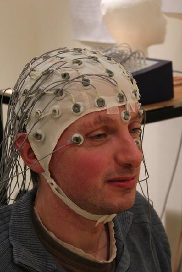
Electroencephalography (EEG) is one technique for studying brain activity. This technique uses at least two and up to 256 electrodes to measure the difference in electrical charge (the voltage) between pairs of points on the head. These electrodes are typically fastened to a flexible cap (similar to a swimming cap) that is placed on the participant’s head. Figure \(\PageIndex{1}\) shows a patient wearing such a cap. From the scalp, the electrodes measure the electrical activity that is naturally occurring within the brain. They do not introduce any new electrical activity.
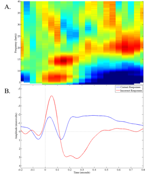
Given that this electrical activity must travel through the skull and scalp before reaching the electrodes, localization of activity is less precise when measuring from the scalp, but it can still be within several millimeters when localizing activity that is near the scalp. While EEG is lacking with respect to spatial resolution, one major advantage of EEG is its temporal resolution. Data can be recorded thousands of times per second, allowing researchers to document events that happen in less than a millisecond. EEG analyses typically investigate the change in amplitude (wave height) or frequency (number of waves per unit of time) components of the recorded EEG on an ongoing basis or averaged over dozens of trials (see Figure \(\PageIndex{2}\)). The EEG has been used extensively in the study of sleep. When you hear references to "brain waves", those are references to information obtained using EEG.
MEG
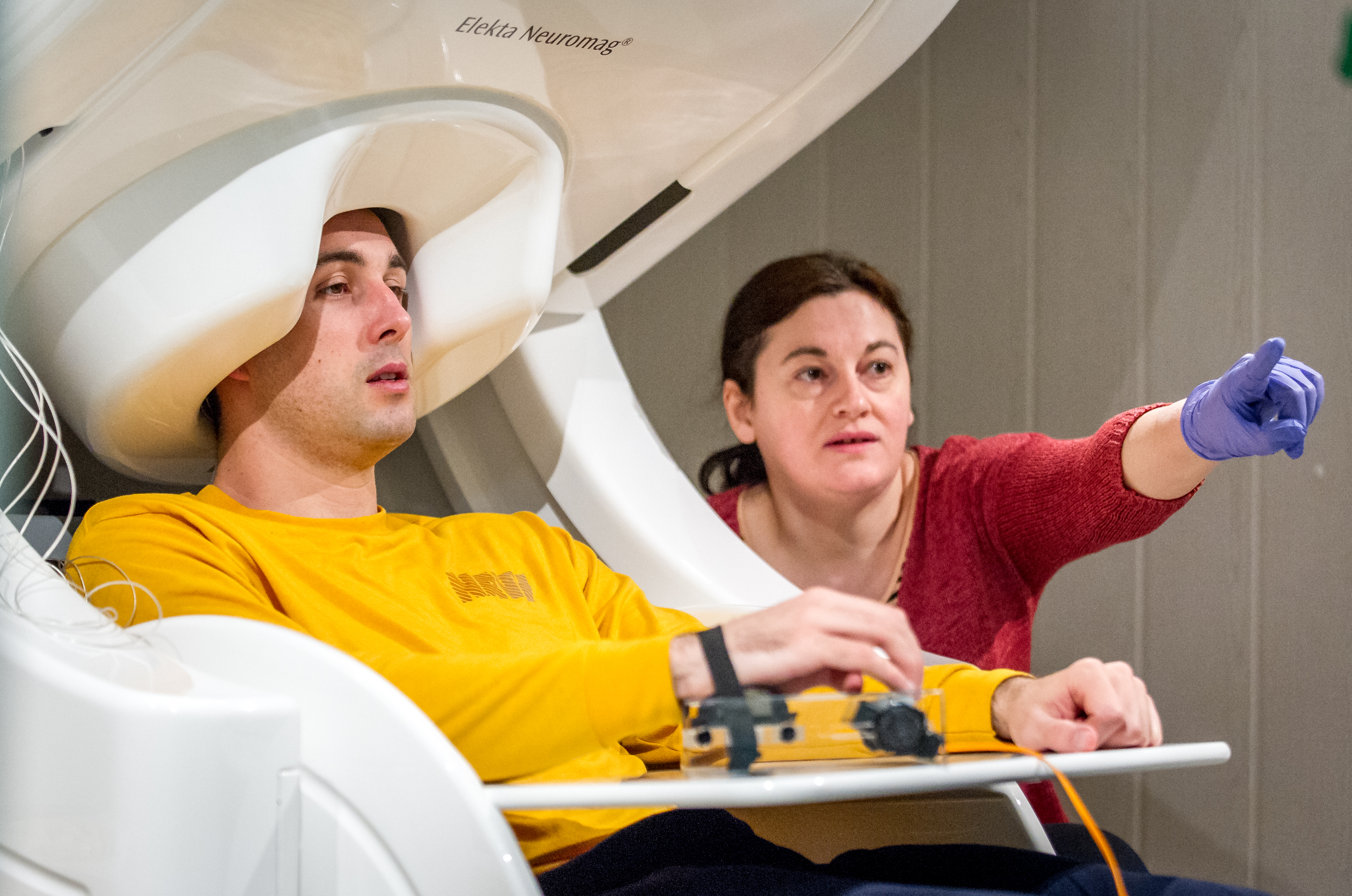
Magnetoencephalography (MEG) is another technique for noninvasively measuring neural activity. The flow of electrical charge (the current) associated with neural activity produces very weak magnetic fields that can be detected by sensors placed near the participant’s scalp. Figure \(\PageIndex{3}\) depicts a subject in an MEG machine. The number of sensors used varies from a few to several hundred. Due to the fact that the magnetic fields of interest are so small, special rooms that are shielded from magnetic fields in the environment are needed in order to avoid contamination of the signal being measured. MEG has the same excellent temporal resolution as EEG. Additionally, MEG is not as susceptible to distortions from the skull and scalp. Magnetic fields are able to pass through the hard and soft tissue relatively unchanged, thus providing better spatial resolution than EEG. MEG analytic strategies are nearly identical to those used in EEG. However, the MEG recording apparatus is much more expensive than EEG, so MEG is much less widely available.
General Information About Direct Imaging Techniques
EEG and MEG both have excellent temporal resolution and are useful when someone is particularly interested in studying the timing of brain activity. For example, if someone is reading a sentence that ends with an unexpected word, how long after reading the unexpected word does the brain react to it? In addition to these types of questions, EEG and MEG methods allow researchers to investigate the degree to which different parts of the brain “talk” to each other. This allows for a better understanding of brain networks, such as their role in different tasks and how they may function abnormally in psychopathology.
Direct imaging techniques are those that allow for a direct measure of brain activity. EEG and MEG are both considered direct brain imaging techniques since EEG measures the electrical activity from groups of neurons and MEG measures the magnetic fields that the electrical activity gives off. Neither of these techniques relies on measuring something else with an assumption that they are linked. This is not true in the next set of techniques we will discuss.
Using Direct Functional Imaging Techniques to Study a Disorder: Autism Spectrum Disorder
EEG and MEG have been used to examine ASD. One of the findings included a delay in the brain wave associated with auditory stimuli. In short, there are differences in the time for processing auditory sounds in children with ASD compared to those without ASD. Furthermore, this delay appears more pronounced in children with ASD who have language developmental delays as opposed to children with ASD without linguistic delays (Roberts et al., 2019). This delay has even been proposed to help clinicians diagnose autism in young children.
Summary
Direct imaging techniques are extremely useful ways to measure electrical brain activity in a non-invasive way. Both EEG and MEG are most useful at identifying differences in timing patterns of electrical activity.
References
Roberts, T. P., Matsuzaki, J., Blaskey, L., Bloy, L., Edgar, J. C., Kim, M., Ku, M., Kuschner, E. S., & Embick, D. (2019). Delayed M50/M100 evoked response component latency in minimally verbal/nonverbal children who have autism spectrum disorder. Molecular Autism, 10(1).
Attributions
- Psychophysiological Methods in Neuroscience by Zachary Infantolino and Gregory A. Miller is licensed under a CC BY-NC-SA 4.0 International License.
- Figure \(\PageIndex{1}\): "EEG Brain Scan" by Tim Sheerman-Chase is licensed under CC BY 2.0
- Figure \(\PageIndex{3}\): "170321-F-LW859-017" by Airman Magazine is licensed under CC BY-NC 2.0