
2.5: Other Non-Invasive Techniques
- Page ID
- 110464
This page is a draft and under active development. Please forward any questions, comments, and/or feedback to the ASCCC OERI (oeri@asccc.org).
- Describe how transcranial magnetic stimulation (TMS) is different from other functional imaging techniques.
- Differentiate between depolarization and hyperpolarization with respect to TMS.
Overview
This section is frequently combined with one of the previous sections in many textbooks because many of the researchers who do functional brain imaging work also conduct TMS work (the subject of this section). However, this technique is different from the previous techniques in many important ways. In this section, we will discuss what TMS is, when it is used, and how it is different from the previous imaging techniques.
Transcranial Magnetic Stimulation
Another technique that is worth mentioning is transcranial magnetic stimulation (TMS). TMS is a noninvasive method that causes depolarization or hyperpolarization in neurons near the scalp. In TMS, a coil of wire is placed just above the participant’s scalp (as shown in Figure \(\PageIndex{1}\)). When electricity flows through the coil, it produces a magnetic field. This magnetic field travels through the skull and scalp and affects neurons near the surface of the brain. When the magnetic field is rapidly turned on and off, a current is induced in the neurons, leading to depolarization or hyperpolarization, depending on the number of magnetic field pulses. Single- or paired-pulse TMS depolarizes site-specific neurons in the cortex, causing them to fire. If this method is used over primary motor cortex, it can produce or block muscle activity, such as inducing a finger twitch or preventing someone from pressing a button. If used over primary visual cortex, it can produce sensations of flashes of light or impair visual processes. This has proved to be a valuable tool in studying the function and timing of specific processes such as the recognition of visual stimuli. Repetitive TMS produces effects that last longer than the initial stimulation. Depending on the intensity, coil orientation, and frequency, neural activity in the stimulated area may be either attenuated or amplified. Used in this manner, TMS is able to explore neural plasticity, which is the ability of connections between neurons to change. This has implications for treating psychological disorders as well as understanding long-term changes in neuronal excitability.
Note that TMS is different from the previous techniques in that we are not taking images of what the brain is doing. TMS disrupts or stimulates the brain and actively changes what the brain is doing.
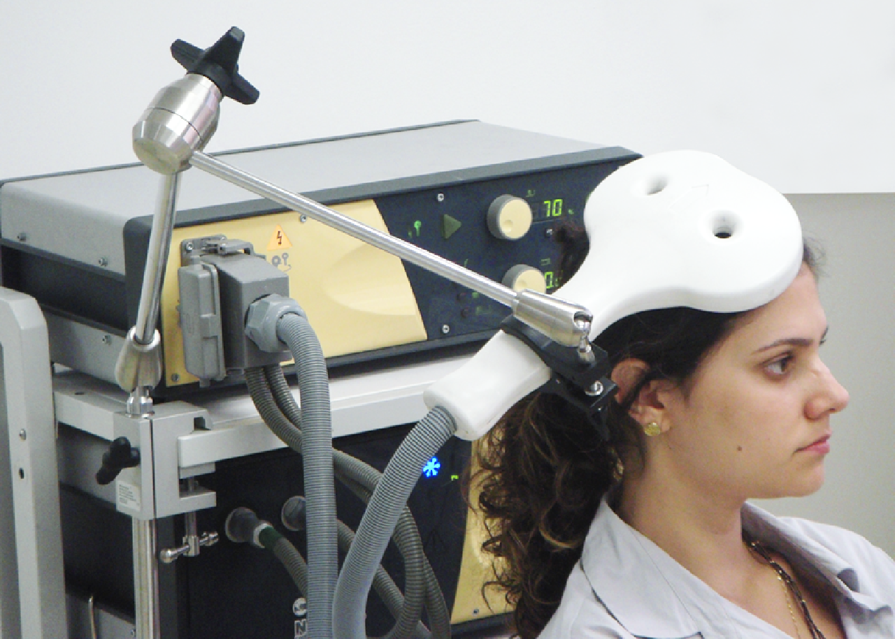
Figure \(\PageIndex{1}\): Woman with TMS wand pressed against the back right side of her head. Credit: "TMS" by Baburov is licensed under CC BY-SA 4.0
Using Transcranial Magnetic Imaging to Study a Disorder: Autism Spectrum Disorder
TMS studies, as with most research techniques, can come in the form of basic research (research intended to inform our understanding) and applied research (research intended to solve a problem). Basic research in neuroscience is typically driven by research questions aimed at a general understanding of how the brain and nervous system work. Some TMS studies have used TMS to reduce brain activity in the right amygdala during the processing of faces with negative emotions (Baeken et al., 2010). Although this research wasn’t specific to autism, it is not hard to see the connection between understanding how the amygdala works and ASD. Furthermore, studies have tried to use TMS to treat ASD. Studies thus far have focused on using TMS to change activity levels and possibly stimulate neural plasticity. There was even a transcranial magnetic stimulation therapy for autism conference held in 2014 to discuss the use of the tool in the treatment of ASD. Indeed, there are myriad of possibilities for how this tool can be used in the future. (See Oberman et al. (2015) for a review of TMS treatments for ASD.)
Summary
TMS is a technique where researchers and doctors are able examine the changes in function that are produced when a brain area is excited or inhibited. For research purposes, it has been useful to help us map the brain. For clinical purposes, TMS is being studied more and more to understand whether it can cause alterations in the neural networks that can lead to benefits for people with various disorders.
References
Baeken, C., De Raedt, R., Van Schuerbeek, P., Vanderhasselt, M. A., De Mey, J., Bossuyt, A., & Luypaert, R. (2010). Right prefrontal HF-rtms attenuates right amygdala processing of negatively valenced emotional stimuli in healthy females. Behavioural Brain Research, 214(2), 450–455.
Oberman, L. M., Enticott, P. G., Casanova, M. F., Rotenberg, A., Pascual-Leone, A., & McCracken, J. T. (2015). Transcranial magnetic stimulation in autism spectrum disorder: Challenges, promise, and roadmap for future research. Autism Research, 9(2), 184–203.
Attributions
- Figure \(\PageIndex{1}\): "TMS" by Baburov is licensed under CC BY-SA 4.0