
9.4: Motor Circuitry- Neural Structures and Pathways
- Page ID
- 116196
This page is a draft and under active development. Please forward any questions, comments, and/or feedback to the ASCCC OERI (oeri@asccc.org).
- Identify the main structures of neural motor circuits at the cortical, subcortical, and spinal levels
- Describe the main functional roles of the basal ganglia, premotor area, and motor cortex
- Apply knowledge of functional cortical anatomy to the control of fine and gross body movements
Neuroanatomy of Motor Systems
The motor system controls all of our skeletal muscle movement and more. There are multiple levels of control. Within the spinal cord, simple reflexes can function without higher input from the brain. Slightly more complex spinal control occurs when central pattern generators function during repetitive movements like walking. The motor, premotor and supplementary cortices in the brain are responsible for the planning and execution of voluntary movements. And finally, the basal ganglia and cerebellum modulate the responses of the neurons in the motor cortex to help with coordination, motor learning, and balance.
Cortical Anatomy of Movement
Much of the cortex is actually involved in the planning of voluntary movement. Sensory information, particularly the dorsal stream of the visual and somatosensory pathways, is processed in the posterior parietal cortex where visual, tactile, and proprioceptive information is integrated.
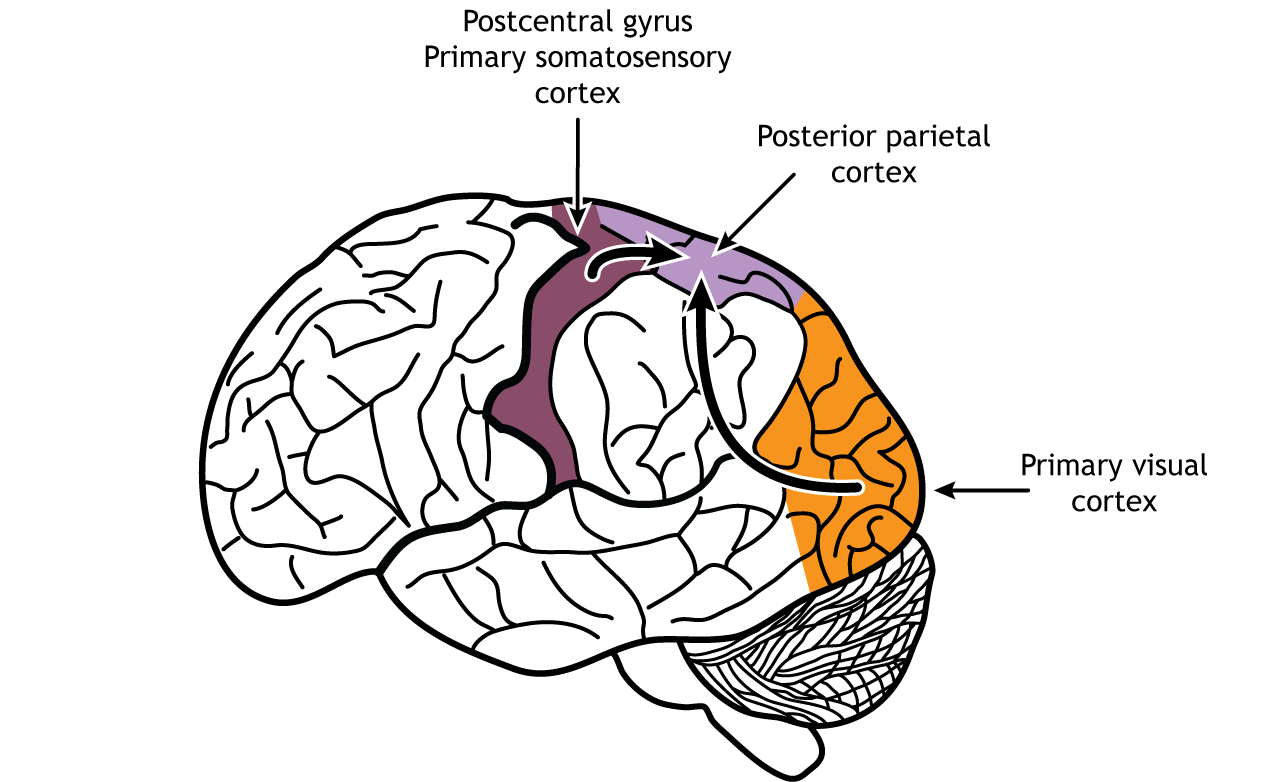
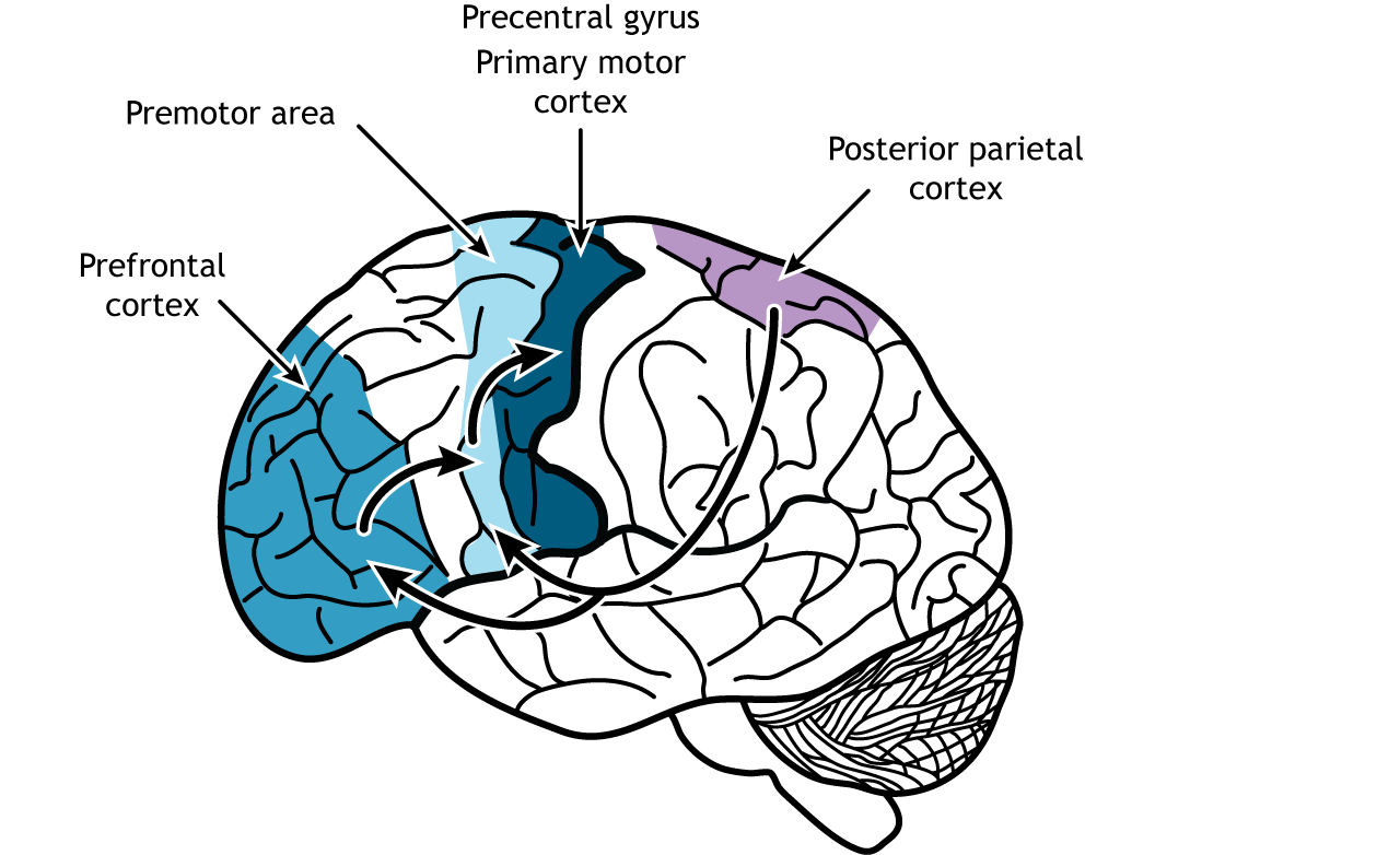
Connections from the posterior parietal cortex are then sent to both the premotor regions and the prefrontal cortex. The prefrontal cortex, which is located in the front of the brain in the frontal lobe, plays an important role in higher level cognitive functions like planning, critical thinking, and understanding the consequences of our behaviors. The premotor area lies just anterior to the primary motor cortex. This region helps plan and organize movement and makes decisions about which actions should be used for a situation.
Role of Premotor Area
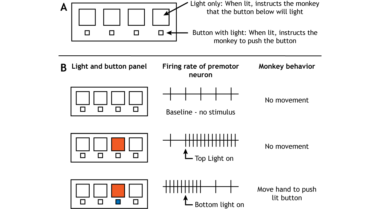
The premotor regions send some axons directly to lower motor neurons in the spinal cord using the same pathways as the motor cortex. However, the premotor cortex also plays an important role in the planning of movement. Two experimental designs have demonstrated this role. Monkeys were trained on a panel that had one set of lights in a row on top and one set of buttons that could also light up in a row on the bottom. The monkeys would watch for a top row light to turn on. This would indicate that within a few seconds, the button directly below would light up. When the button turned on, the monkeys were supposed to push the button.
Therefore, there were two light triggers in the experiment. The first required no motor movement from the monkey but did give the monkey information about where a motor movement would be needed in the near future. The second required the monkey to move to push the button. When brain activity was measured during this study, neurons in the premotor cortex became active when the first light trigger turned on, well before any movement actually took place (Weinrich and Wise, 1928).
In another experiment, people were trained to move their fingers in a specific pattern. Cerebral blood flow was then measured when they repeated the finger pattern and when they only imagined repeating the finger pattern. When the movement was only imagined and not actually executed, the premotor regions along with parts of the prefrontal cortex were activated (Roland, et al, 1980).
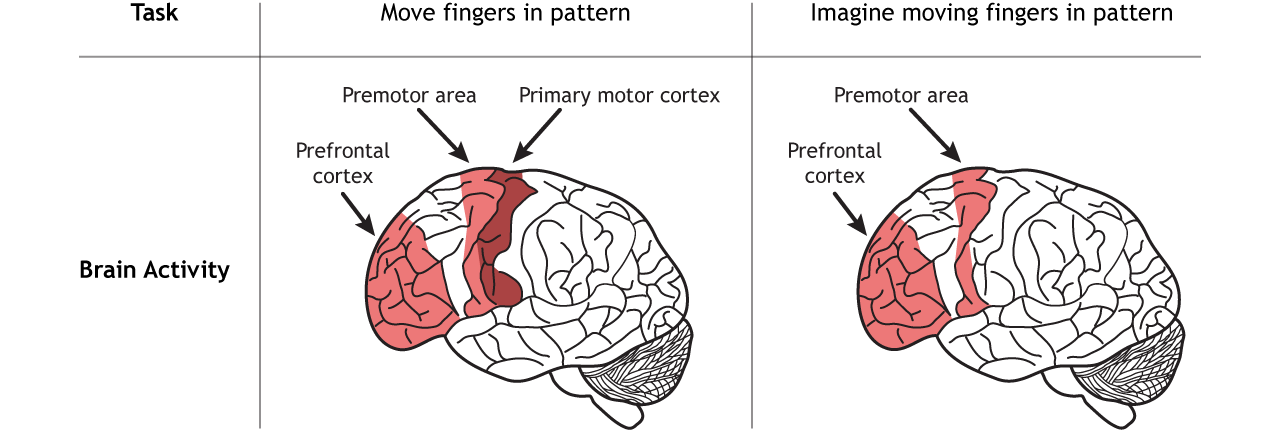
These studies show that the premotor cortex is active prior to the execution of movement, indicating that it plays an important role in the planning of movement. The posterior parietal, prefrontal, and premotor regions, though, also communicate with a subcortical region called the basal ganglia to fully construct the movement plan. The basal ganglia are covered in the next subsection.
Role of Motor Cortex
Once the plan for movement has been created, the primary motor cortex is responsible for the execution of that action. The primary motor cortex lies just anterior to the primary somatosensory cortex in the precentral gyrus located in the frontal lobe.
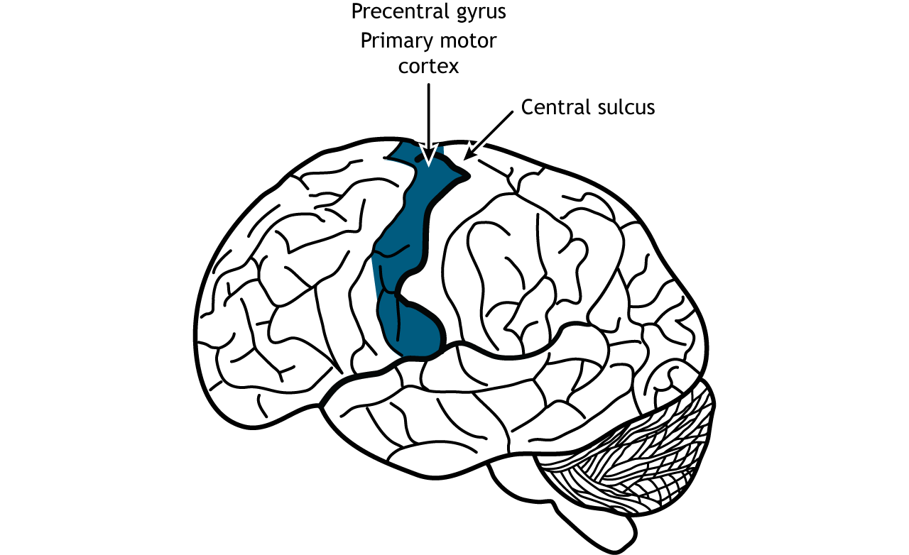
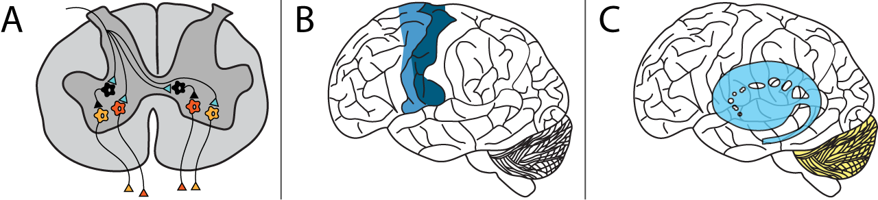
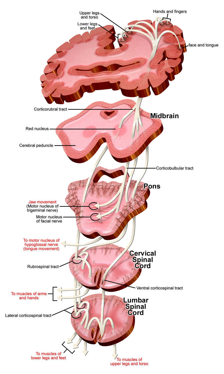
Like the somatosensory cortex, the motor cortex is organized by a somatotopic map in that different areas of the body are controlled by distinct areas of the motor cortex. However, the motor cortex does not map onto the body in such an exact way as does the somatosensory system. It is believed that upper motor neurons in the motor cortex control multiple lower motor neurons in the spinal cord that innervate multiple muscles. This results in activation of an upper motor neuron causing excitation or inhibition in different neurons at once, indicating that the primary motor cortex is responsible for movements and not simply activation of one muscle. Stimulation of motor neurons in monkeys can lead to complex motions like bringing the hand to the mouth or moving into a defensive position (Graziano et al, 2005).
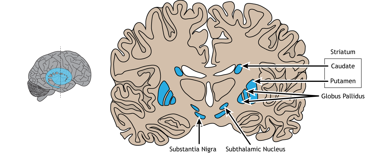
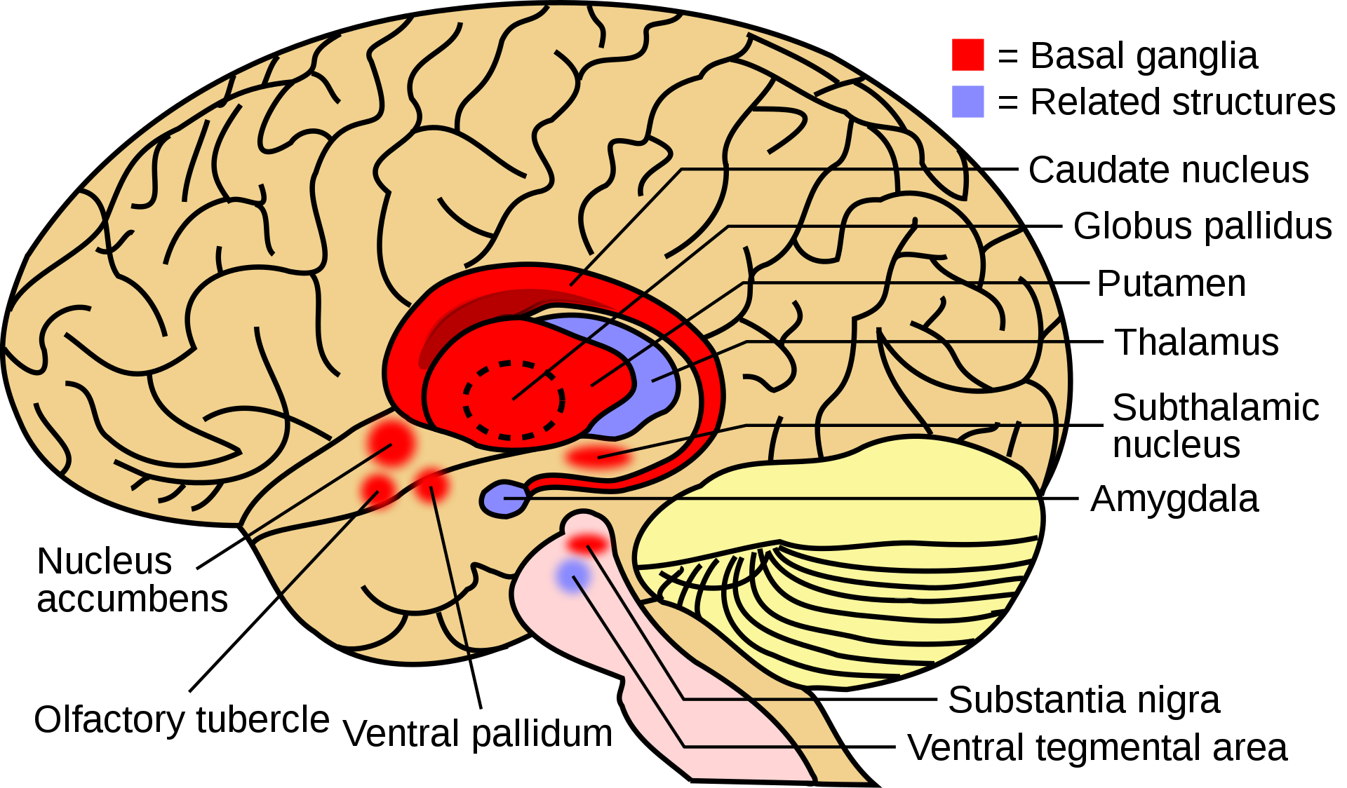
Basal Ganglia
The basal ganglia are a group of subcortical nuclei, meaning groups of neurons that lie below the cerebral cortex. The basal ganglia is comprised of several nuclei including the caudate nucleus, putamen, and the globus pallidus. Another nucleus typically associated with the basal ganglia is the subthalamic nucleus. The substantia nigra of the midbrain provides dopaminergic input to these areas.
The basal ganglia are primarily associated with motor control, since motor disorders, such as Parkinson’s or Huntington’s diseases stem from dysfunction of neurons within the basal ganglia. For voluntary motor behavior, the basal ganglia are involved in the initiation or suppression of behavior and can regulate movement through modulating activity in the thalamus and cortex. In addition to motor control, the basal ganglia also communicate with non-motor regions of the cerebral cortex and play a role in other behaviors such as emotional and cognitive processing.
Basal Ganglia Input
The majority of information processed by the basal ganglia enters through the striatum. The principal source of input to the basal ganglia is from the cerebral cortex. This input is glutamatergic (i.e. uses glutamate as its neurotransmitter) and therefore, excitatory. The substantia nigra is also a region with critical projections to the striatum and is the main source of dopaminergic input. Dopamine plays an important role in basal ganglia function. Parkinson’s disease results when dopamine neurons in the substantia nigra degenerate and no longer send appropriate inputs to the striatum. Dopamine projections can have either excitatory or inhibitory effects in the striatum, depending on the type of metabotropic dopamine receptor the striatal neuron expresses. Dopamine action at a neuron that expresses the D1 receptor is excitatory. Dopamine action at a neuron that expresses the D2 receptor is inhibitory.
Basal Ganglia Output
The primary output region of the basal ganglia is the internal segment of the globus pallidus. This region sends inhibitory GABAergic projections to nuclei in the thalamus. This inhibitory output has a tonic, constant firing rate, which allows the basal ganglia output to both increase and decrease depending on the situation. The thalamus then projects back out to the cerebral cortex, primarily to motor areas.
Attributions:
- Adapted from Foundations of Neuroscience - Chapter 26. Authored by Casey Henley is licensed under a License: (CC BY-NC-SA) 4.0 International License. https://openbooks.lib.msu.edu/neuros...g-of-movement/