This section will explain where and how language is processed. To avoid intersections with visual processes we will firstly concentrate on spoken language. Scientists have developed three approaches of conceiving information about this issue. The first two approaches are based upon brain lesions, namely aphasia, whereas the recent approach relies on results of on modern brain-image techniques.
Neurological Perspective
The Neurological Perspective describes which pathways language follows in order to be comprehended. Scientists revealed that there are concrete areas inside the brain where concrete tasks of language processing are taking place. The most known areas are the Broca and the Wernicke Area.
Broca’s aphasia
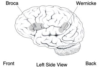
Broca's and Wernicke's area
One of the most well-known aphasias is Broca’s aphasia that causes patients to be unable to speak fluently. Moreover they have a great difficulty producing words. Comprehension, however, is relatively intact in those patients. Because these symptoms do not result from motoric problems of the vocal musculature, a region in the brain that is responsible for linguistic output must be lesioned. Broca discovered that the brain region causing fluent speech is responsible for linguistic output, must be located ventrally in the frontal lobe, anterior to the motor strip. Recent research suggested that Broca`s aphasia results also from subcortical tissue and white matter and not only cortical tissue.
| Example of spontaneous Speech - Task: What do you see on this picture? |
| „O, yea. Det‘s a boy an‘ girl... an‘ ... a ... car ... house... light po‘ (pole). Dog an‘ a ... boat. ‚N det‘s a ... mm ... a ... coffee, an‘ reading. Det‘s a ... mm ... a ... det‘s a boy ... fishin‘.“ (Adapted from „Principles of Neuroscience“ 4th edition, 2000, p 1178) |
Wernicke‘s aphasia
Another very famous aphasia, known as Wernicke`s aphasia, causes opposite syndromes. Patients suffering from Wernicke`s aphasia usually speak very fluently, words are pronounced correctly, but they are combined senselessly – “word salad” is the way it is most often described. Understanding what patients of Wernicke`s aphasia say is especially difficult, because they use paraphasias (substitution of a word in verbal paraphasia, of word with similar meaning in semantic paraphasia, and of a phoneme in phonemic paraphasia) and neologisms. With Wernicke`s aphasia the comprehension of simple sentences is a very difficult task. Moreover their ability to process auditory language input and also written language is impaired. With some knowledge about the brainstructure and their tasks one is able to conclude that the area that causes Wernicke`s aphasia, is situated at the joint of temporal, parietal and occipital regions, near Heschl`s gyrus (primary auditory area), because all the areas receiving and interpreting sensory information (posterior cortex), and those connecting the sensory information to meaning (parietal lobe) are likely to be involved.
| Example of spontaneous Speech - Task: What do you see on this picture? |
| „Ah, yes, it‘s ah ... several things. It‘s a girl ... uncurl ... on a boat. A dog ... ‘S is another dog ... uh-oh ... long‘s ... on a boat. The lady, it‘s a young lady. An‘ a man a They were eatin‘. ‘S be place there. This ... a tree! A boat. No, this is a ... It‘s a house. Over in here ... a cake. An‘ it‘s, it‘s a lot of water. Ah, all right. I think I mentioned about that boat. I noticed a boat being there. I did mention that before ... Several things down, different things down ... a bat ... a cake ... you have a ...“ (adapted from „Principles of Neuroscience“ 4th edition, 2000, p 1178) |
Conduction aphasia
Wernicke supposed that an aphasia between Broca‘s area and Wernicke‘s area, namely conduction aphasia, would lead to severe problems to repeat just heard sentences rather than having problems with the comprehension and production of speech. Indeed patients suffering from this kind of aphasia show an inability to reproduce sentences since they often make phonemic paraphasias, may substitute or leave out words, or might say nothing. Investigations determined that the "connection cable", namely the arcuate fasciculus between Wernicke‘s and Broca‘s area is almost invariably damaged in case of a conduction aphasia. That is why conduction aphasia is also regarded as a disconnection syndrome (the behavioural dysfunction because of a damage to the connection of two connected brain regions).
| Example of the repetition of the sentence „The pastry-cook was elated“: |
| „The baker-er was /vaskerin/ ... uh ...“ (adapted from „Principles of Neuroscience“ 4th edition, 2000, p 1178) |
Transcortical motor aphasia and global aphasia
Transcortical motor aphasia, another brain lesion caused by a connection disruption, is very similar to Broca`s aphasia, with the difference that the ability to repeat is kept. In fact people with a transcortical motor aphasia often suffer from echolalia, the need to repeat what they just heard. Usually patients` brain is damaged outside Broca`s area, sometimes more anterior and sometimes more superior. Individuals with transcortical sensory aphasia have similar symptoms as those suffering from Wernicke`s aphasia, except that they show signs of echolalia. Lesions in great parts of the left hemisphere lead to global aphasia, and thus to an inability of both comprehending and producing language, because not only Broca`s or Wenicke`s area is damaged. (Barnich, 1997, pp. 276–282)
Overview of the effects of aphasia from the neurological perspective
| Type of Aphasia |
Spontaneous Speech |
Paraphasia |
Comprehension |
Repetition |
Naming |
- Broca`s
- Wernicke`s
- Conduction
- Transcortical motor
- Transcortical sensory
- Global
|
- Nonfluent
- Fluent
- Fluent
- Nonfluent
- Fluent
- Nonfluent
|
- Uncommon
- Common (verbal)
- Common (literal)
- Uncommon
- Common
- Variable
|
- Good
- Poor
- Good
- Good
- Poor
- Poor
|
- Poor
- Poor
- Poor
- Good (echolalia)
- Good (echolalia)
- Poor
|
- Poor
- Poor
- Poor
- Poor
- Poor
- Poor
|
(Adapted from Benson, 1985,p. 32 as cited in Barnich, 1997, p. 287)
Psychological Perspective
Since the 1960‘s psychologists and psycholinguists tried to resolve how language is organised and represented inside the brain. Patients with aphasias gave good evidence for location and discrimination of the three main parts of language comprehension and production, namely phonology, syntax and semantics.
Phonology
Phonology deals with the processing of meaningful parts of speech resulting from the mere sound. More over there exists a differentiation between a phonemic representation of a speech sound which are the smallest units of sounds that leads to different meanings (e.g. the /b/ and /p/ in bet and pat) and phonetic representation. The latter means that a speech sound may be produced in a different manner at different situations. For instance the /p/ in pill sounds different than the /p/ in spill since the former /p/ is aspirated and the latter is not.
Examining which parts are responsible for phonetic representation, patients with Broca`s or Wernicke`s aphasia can be compared. As the speech characteristic for patients with Broca`s aphasia is non-fluent, i.e. they have problems producing the correct phonetic and phonemic representation of a sound, and people with Wernicke`s aphasia do not show any problems speaking fluently, but also have problems producing the right phoneme. This indicates that Broca`s area is mainly involved in phonological production and also, that phonemic and phonetic representation do not take place in the same part of the brain. Scientists examined on a more precise level the speech production, on the level of the distinctive features of phonemes, to see in which features patients with aphasia made mistakes.
A distinctive feature describes the different manners and places of articulation. /t/ (like in touch) and /s/ (like in such) for example are created at the same place but produced in different manner. /t/ and /d/ are created at the same place and in the same manner but they differ in voicing.
Results show that in fluent as well as in non-fluent aphasia patients usually mix up only one distinctive feature, not two. In general it can be said that errors connected to the place of articulation are more common than those linked to voicing. Interestingly some aphasia patients are well aware of the different features of two phonemes, yet they are unable to produce the right sound. This suggests that though patients have great difficulty pronouncing words correctly, their comprehension of words is still quite good. This is characteristic for patients with Broca`s aphasia, while those with Wernicke`s aphasia show contrary symptoms: they are able to pronounce words correctly, but cannot understand what the words mean. That is why they often utter phonologically correct words (neologisms) that are not real words with a meaning.
Syntax
Syntax describes the rules of how words must be arranged to result in meaningful sentences. Humans in general usually know the syntax of their mother tongue and thus slip their tongue if a word happens to be out of order in a sentence. People with aphasia, however, often have problems with parsing of sentences, not only with respect to the production of language but also with respect to comprehension of sentences. Patients showing an inability of comprehension and production of sentences usually have some kind of anterior aphasia, also called agrammatical aphasia. This can be revealed in tests with sentences. These patients cannot distinguish between active and passive voice easily if both agent and object could play an active part. For example patients do not see a difference between “The boy chased the girl” and “The boy was chased by the girl”, but they do understand both “The boy saw the apple” and “The apple was seen by the boy”, because they can seek help of semantics and do not have to rely on syntax alone. Patients with posterior aphasia, like for example Wernicke`s aphasia, do not show these symptoms, as their speech is fluent. Comprehension by mere syntactic means would be possible as well, but the semantic aspect must be considered as well. This will be discussed in the next part.
Semantics
Semantics deals with the meaning of words and sentences. It has been shown that patients suffering from posterior aphasia have severe problems understanding simple texts, although their knowledge of syntax is intact. The semantic shortcoming is often examined by a Token Test, a test in which patients have to point to objects referred to in simple sentences. As might have been guessed, people with anterior aphasia have no problems with semantics, yet they might not be able to understand longer sentences because the knowledge of syntax then is involved as well.
Overview of the effects of aphasia from the psychological perspective
| |
anterior Aphasia (e.g. Broca) |
posterior Aphasia (e.g. Wernicke) |
| Phonology |
phonetic and phonemic representation affected |
phonemic representation affected |
| Syntax |
affected |
no effect |
| Syntax |
no effect |
affected |
In general studies with lesioned people have shown that anterior areas are needed for speech output and posterior regions for speech comprehension. As mentioned above anterior regions are also more important for syntactic processing, while posterior regions are involved in semantic processing. But such a strict division of the parts of the brain and their responsibilities is not possible, because posterior regions must be important for more than just sentence comprehension, as patients with lesions in this area can neither comprehend nor produce any speech. (Barnich, 1997, pp. 283–293)
Evidence from Advanced Neuroscience Methods
Measuring the functions of both normal and damaged brains has been possible since the 1970s, when the first brain imaging techniques were developed. With them, we are able to “watch the brain working” while the subject is e.g. listening to a joke. These methods (further described in chapter 4) show whether the earlier findings are correct and precise.
Generally, imaging shows that certain functional brain regions are much smaller than estimated in brain lesion studies, and that their boundaries are more distinct (cf. Banich p. 294). The exact location varies individually, therefore bringing the results of many brain lesion studies together caused too big estimated functional regions before. For example, stimulating brain tissue electrically (during epilepsy surgery) and observing the outcome (e.g. errors in naming tasks) led to a much better knowledge where language processing areas are located.
PET studies (Fiez & Petersen, 1993, as cited in Banich, p. 295) have shown that in fact both anterior and posterior regions were activated in language comprehension and processing, but with different strengths – in agreement with the lesion studies. The more active speech production is required in experiments, the more frontal is the main activation: For example, when the presented words must be repeated.
Another result (Raichle et al. 1994, as referred to in Banich, p. 295) was that the familiarity of the stimuli plays a big role. When the subjects were presented well-known stimuli sets in well-known experimental tasks and had to repeat them, anterior regions were activated. Those regions were known to cause conduction aphasia when damaged. But when the words were new ones, and/or the subjects never had to do a task like this before, the activation was recorded more posterior. That means, when you repeat an unexpected word, the heaviest working brain tissue is about somewhere under your upper left earlap, but when you knew this word that would be the next to repeat before, it is a bit nearer to your left eye.

