
3.1.2: Introduction to Genetics
- Last updated
- Save as PDF
- Page ID
- 136385
Genetics is the study of heredity. Parents pass down their genetic traits to their offspring. Although children resemble their parents, traits often vary in appearance or molecular function. For example, two parents with normal color vision can sometimes produce a son with red-green colorblindness. Patterns of genetic inheritance will be discussed in a later section. Molecular geneticists study the biological mechanisms responsible for creating variation between individuals, such as DNA mutations (see Chapter 4), cell division, and genetic regulation.
Molecular anthropologists use genetic data to test anthropological questions. Although their interests are diverse, areas of molecular anthropology research include the following: human origins, dispersals, evolution, adaptation, demography, health, disease, behavior, and animal domestication. In addition to conducting research in a laboratory, molecular anthropologists also work in the field with different communities of people. Some anthropologists also study DNA from individuals who have been deceased for decades—even hundreds or thousands of years. The study of has led to the development of specialized laboratory techniques. Over time, the DNA in skeletons of ancient individuals becomes degraded (i.e., less intact), which is why careful methodological considerations must be taken. A recent example of an aDNA study is provided in Special Topic: Native American Immunity and European Diseases, and another will be presented in Chapter 10.
SPECIAL TOPIC: FOCUS ON NATIVE AMERICAN IMMUNITY AND EUROPEAN DISEASES—A STUDY OF ANCIENT DNA
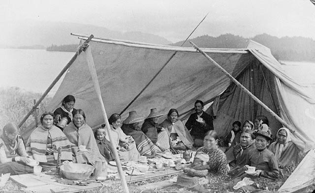 Figure \(\PageIndex{1}\): Tsimshian Native Americans of the Pacific Northwest Coast.
Beginning in the early 15th century, Native Americans progressively suffered from high mortality rates as the result of colonization from foreign powers. European-borne diseases such as measles, tuberculosis, influenza, and smallpox are largely responsible for the population collapse of indigenous peoples in the Americas. Many Europeans who immigrated to the New World had lived in large sedentary populations, which also included coexisting with domestic animals and pests. Although a few prehistoric Native American populations can be characterized as large agricultural societies (especially in Mesoamerica), their overall culture, community lifestyle, and subsistence practices were markedly different from that of Europeans. Therefore, because they did not share the same urban living environments as Europeans, it is believed that Native Americans were susceptible to old-world diseases.
 Figure \(\PageIndex{2}\): Tsimshian territory in present-day British Columbia.
In 2016, a Nature article published by John Lindo and colleagues was the first to investigate whether pre-contact Native Americans possessed a genetic susceptibility to European diseases. Their study included Tsimshians, a First Nation community from British Columbia (Figure 3.12). The DNA from both present-day and ancient individuals (who lived between 500 and 6,000 years ago) was analyzed. The research team discovered that a change occurred in the genetic region HLADQ-1, which is a member of the major histocompatibility complex (MHC) immune system molecules. These molecules are responsible for detecting and triggering an immune response against pathogens. Lindo and colleagues (2016) concluded that HLADQ-1 helped Native Americans adapt to their local environmental ecology. However, when European-borne epidemics occurred in the Northwest during the 1800s, a certain HLADQ-1 DNA sequence associated with ancient Tsimshian immunity was no longer adaptive. As the result of past selective pressures from European diseases, present-day Tsimshians have a different frequency of HLADQ-1 sequences. The precise role that HLADQ-1 plays in immune adaptation still requires further investigation. But overall, this study serves as an example of how studying ancient DNA from the remains of deceased individuals can help provide insight into living human populations and historical events.
DNA Carries Hereditary Information
Surprisingly, the study of inheritance preceded the discovery of DNA. For a period of time, it was believed that proteins carried the hereditary information passed from parents to offspring. Then, in 1944, Oswald Avery, Colin MacLeod, and Maclyn McCarty discovered an association between extracted nucleic acids and the success of their bacterial genetic experiments. Specifically, they demonstrated that DNA was the molecule responsible for the genetic transformation of their pneumonia bacterial strains. Although this was revolutionary work at the time, the field of molecular biology did not fully embrace their findings (it has also been suggested that they were overlooked for a Nobel Prize). It was eventually accepted by the scientific community that DNA is the hereditary material of an organism, especially after the chemical structure of DNA was revealed.
DNA Structure
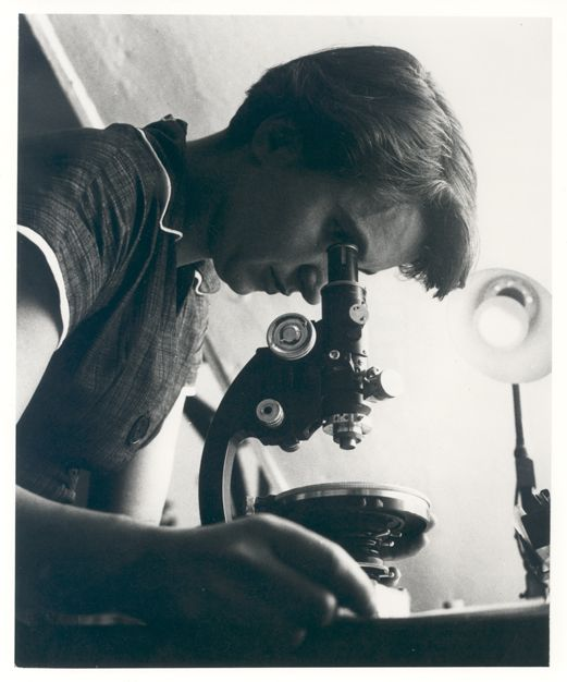 Figure \(\PageIndex{3}\): Chemist and X-ray crystallographer Rosalind Franklin.
The 1953 discovery of the molecular structure of DNA was one of the greatest scientific achievements of all time. Using X-ray crystallography, Rosalind Franklin (Figure 3.13) provided the image that clearly showed the double helix shape of DNA. However, due to a great deal of controversy, Franklin’s colleague and outside associates received greater publicity for the discovery. In 1962, James Watson, Francis Crick, and Maurice Wilkins received a Nobel Prize for developing a biochemical model of DNA. Unfortunately, Rosalind Franklin had passed away in 1958 from ovarian cancer. In current times, Franklin’s important contribution and her reputation as a skilled scientist are widely acknowledged.
The double helix shape of DNA can be described as a twisted ladder (refer back to Figure 3.2). More specifically, DNA is a double-stranded molecule with its two strands oriented in opposite directions (i.e., antiparallel). Each strand is composed of nucleotides with a sugar phosphate backbone. There are four different types of DNA nucleotides: adenine (A), thymine (T), cytosine (C), and guanine (G). The two DNA strands are held together by nucleotide base pairs, which have chemical bonding rules. The complementary base-pairing rules are as follows: A and T bond with each other, while C and G form a bond. The chemical bonds between A—T and C—G are formed by “weak” hydrogen atom interactions, which means the two strands can be easily separated. A DNA sequence is the order of nucleotide bases (A, T, G, C) along only one DNA strand. If one DNA strand has the sequence CATGCT, then the other strand will have a complementary sequence GTACGA. This is an example of a short DNA sequence. In reality, there are approximately three billion DNA base pairs in human cells.
DNA Is Highly Organized Within the Nucleus
If you removed the DNA from a single human cell and stretched it out completely, it would measure approximately two meters (about 6.5 feet). Therefore, DNA molecules must be compactly organized in the nucleus. To achieve this, the double helix configuration of DNA undergoes coiling. An analogy would be twisting a string until coils are formed and then continuing to twist so that secondary coils are formed, and so on. To assist with coiling, DNA is first wrapped around proteins called histones. This creates a complex called chromatin, which resembles “beads on a string” (Figure 3.14). Next, chromatin is further coiled into a chromosome. Another important feature of DNA is that chromosomes can be altered from tightly coiled (chromatin) to loosely coiled (euchromatin). Most of the time, chromosomes in the nucleus remain in a euchromatin state so that DNA sequences are accessible for regulatory processes to occur.
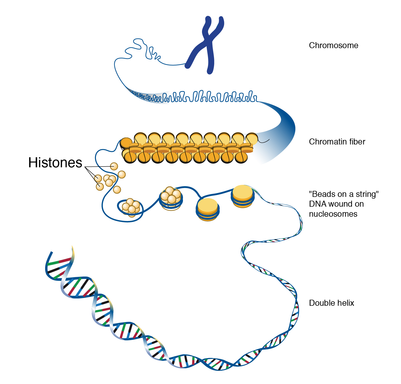 Figure \(\PageIndex{4}\): The hierarchical organization of chromosomes.
Human body cells typically have 23 pairs of chromosomes, for a total of 46 chromosomes in each cell’s nucleus (Figure 3.15). An interesting fact is that the number of chromosomes an organism possesses varies, and this figure is not dependent upon the size or complexity of the organism. For instance, chimpanzees have a total of 48 chromosomes, while hermit crabs have 254. Chromosomes also have a distinct physical structure, including centromeres (the “centers”) and telomeres (the ends) (Figure 3.16). Because of centromeres, chromosomes are described as having two different “arms,” where one arm is long and the other is shorter. Centromeres play an important role during cell division, which will be discussed in the next section. Telomeres are located at the ends of chromosomes and they help protect the chromosomes from degradation after every round of cell division. However, our telomeres become shorter as we age, and if chromosome telomeres become too short, then the cell will stop dividing. Therefore, the link between the regulation of telomere length and cellular aging is of great interest to researchers.
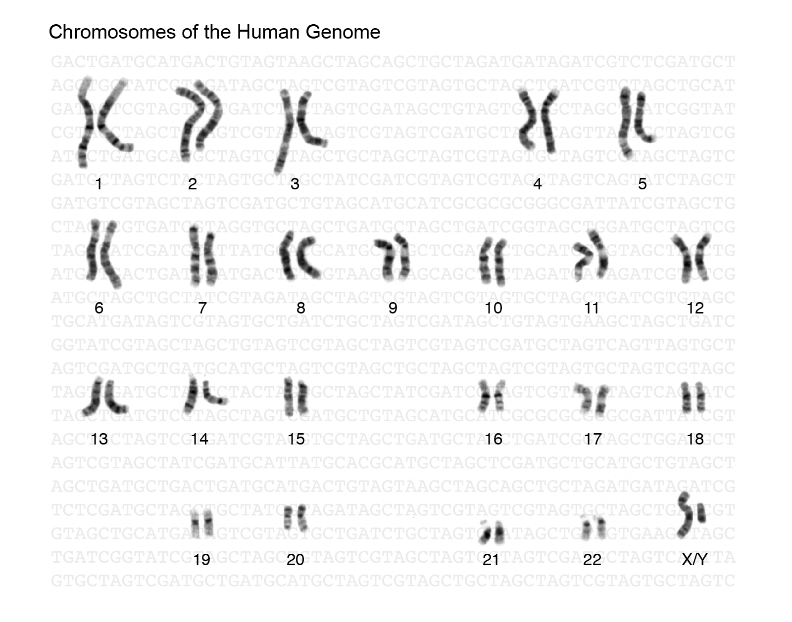 Figure \(\PageIndex{5}\): The 23 human chromosome pairs.
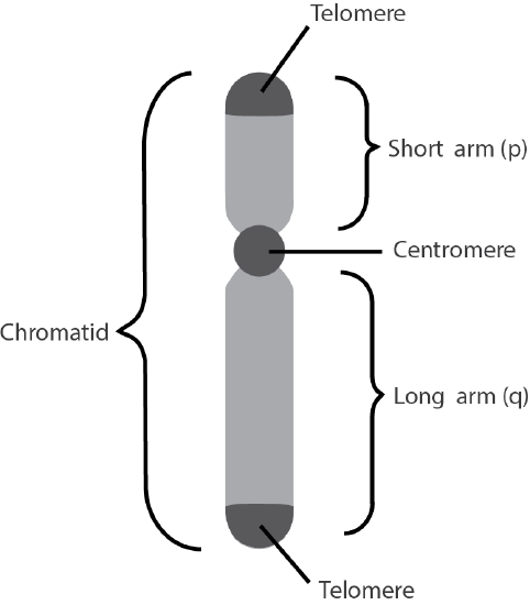 Figure \(\PageIndex{6}\):The regions of a chromosome.
DNA REPLICATION AND CELL DIVISION
For life to continue and flourish, cells must be able to divide. Tissue growth and cellular damage repair are also necessary to maintain an organism throughout its life. All these rely on the dynamic processes of DNA replication and the cell cycle. The mechanisms highlighted in this section are tightly regulated and represent only part of the life cycle of a cell.
DNA Replication
DNA replication is the process by which new DNA is copied from an original DNA template. It is one phase of the highly coordinated cell cycle and requires a variety of enzymes with special functions. Specifically, enzymes carry out the structural and high-energy reactions associated with replicating a double helical molecule. The creation of a complementary DNA strand from a template strand is described as . The result of semi-conservative replication is two separate double-stranded DNA molecules, each of which is composed of an original “parent” template strand and a newly synthesized “daughter” DNA strand.
DNA replication progresses in three steps referred to as initiation, elongation, and termination. Initiation denotes the start of DNA replication by recruiting enzymes to specific sites along the DNA sequence. For example, the double helix of DNA presents structural challenges for replication, so an initiator enzyme, called helicase, “unwinds” DNA by breaking the hydrogen bonds between the two parent strands. The unraveling of the helix into two separated strands creates a fork, which is the active site of replication machinery (Figure 3.17). Once both strands are separated, the parent template strands are exposed, meaning they can be read and replicated.
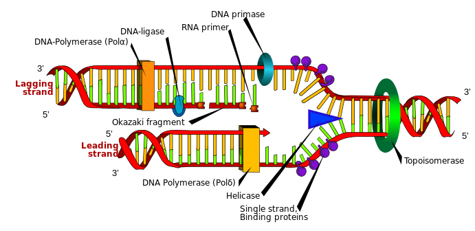 Figure \(\PageIndex{7}\): The different enzymes associated with DNA replication.
Elongation describes the assembly of new DNA daughter strands from parent strands. The two parent strands can further be classified as leading strand or lagging strand and are distinguished by the continuous or discontinuous direction of replication, respectively. A short fragment of RNA nucleotides acts as a primer, which binds to the parent DNA strand that will be copied. The leading strand receives one primer and the lagging strand receives several. Elongation proceeds with help from enzymes called DNA polymerases, which read parent template strands in a specific direction. Complementary nucleotides are added, and the newly formed daughter strand will grow. The direction in which replication proceeds depends on whether it is the leading or lagging strand. On the leading parent strand, a DNA polymerase will create one continuous strand. Because the lagging parent strand requires several primers, disjointed strands (called Okazaki fragments) will be generated. Other enzymes will fill in the missing nucleotide gaps between the disconnected lagging strand Okazaki fragments.
Finally, termination refers to the end of DNA replication activity. It is signaled by a stop sequence in the DNA, which is recognized by machinery at the replication fork. The end result of DNA replication is that the number of chromosomes are doubled so that the cell can divide into two.
DNA Mutations
DNA replication should result in the creation of two molecules with identical DNA nucleotide sequences. Although DNA polymerases are quite precise during DNA replication, copying mistakes are estimated to occur every 107 DNA nucleotides. Variation from the original DNA sequence is known as a mutation. The different types of mutations will be discussed in greater detail in Chapter 4. Briefly, mutations can result in single nucleotide changes as well as the insertion or deletion of nucleotides and repeated sequences. Depending on where they occur, mutations can be deleterious (harmful). For example, mutations may occur in regions that control cell cycle regulation, which can result in cancer (see Special Topic: The Cell Cycle and Immortality of Cancer Cells). Many other mutations, however, are not harmful to an organism.
Regardless of their effect, the cell attempts to reduce the frequency of mutations that occur during DNA replication. To accomplish this, there are polymerases with proofreading capacities that can identify and correct mismatched nucleotides. These safeguards reduce the frequency of DNA mutations so that they only occur every 109 nucleotides.
SPECIAL TOPIC: THE CELL CYCLE AND IMMORTALITY OF CANCER CELLS
DNA replication is part of a series of preparatory phases that a cell undergoes prior to cell division, collectively known as interphase (Figure 3.18). During interphase, the cell not only doubles its chromosomes through DNA replication, but it also increases its metabolic capacity to provide energy for growth and division. Transition into each phase of the cell cycle is tightly controlled by proteins that serve as checkpoints. If a cell fails to pass a checkpoint, then DNA replication and/or cell division will not continue. Some of the reasons why a cell may fail at a checkpoint is DNA damage, lack of nutrients to continue the process, or insufficient size. In turn, a cell may undergo apoptosis, which is a mechanism for cell death.
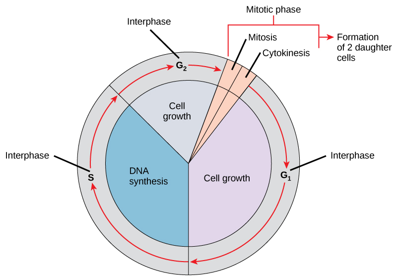 Figure \(\PageIndex{8}\): The phases and checkpoints of the cell cycle.
Unchecked cellular growth is a distinguishing hallmark of cancer. In other words, as cancer cells grow and proliferate, they acquire the capacity to avoid death and replicate indefinitely. This uncontrolled and continuous cell division is also known as “immortality.” As previously discussed, most cells lose the ability to divide due to shortening of telomeres on the ends of chromosomes over time. One way in which cancer cells retain replicative immortality is that the length of their telomeres is continuously protected. Chemotherapy is often used to treat cancer by targeting cell division, which halts the propagation of genetically abnormal cells. Another therapeutic approach that continues to be investigated is targeting telomere activity to stop the division of cancer cells.
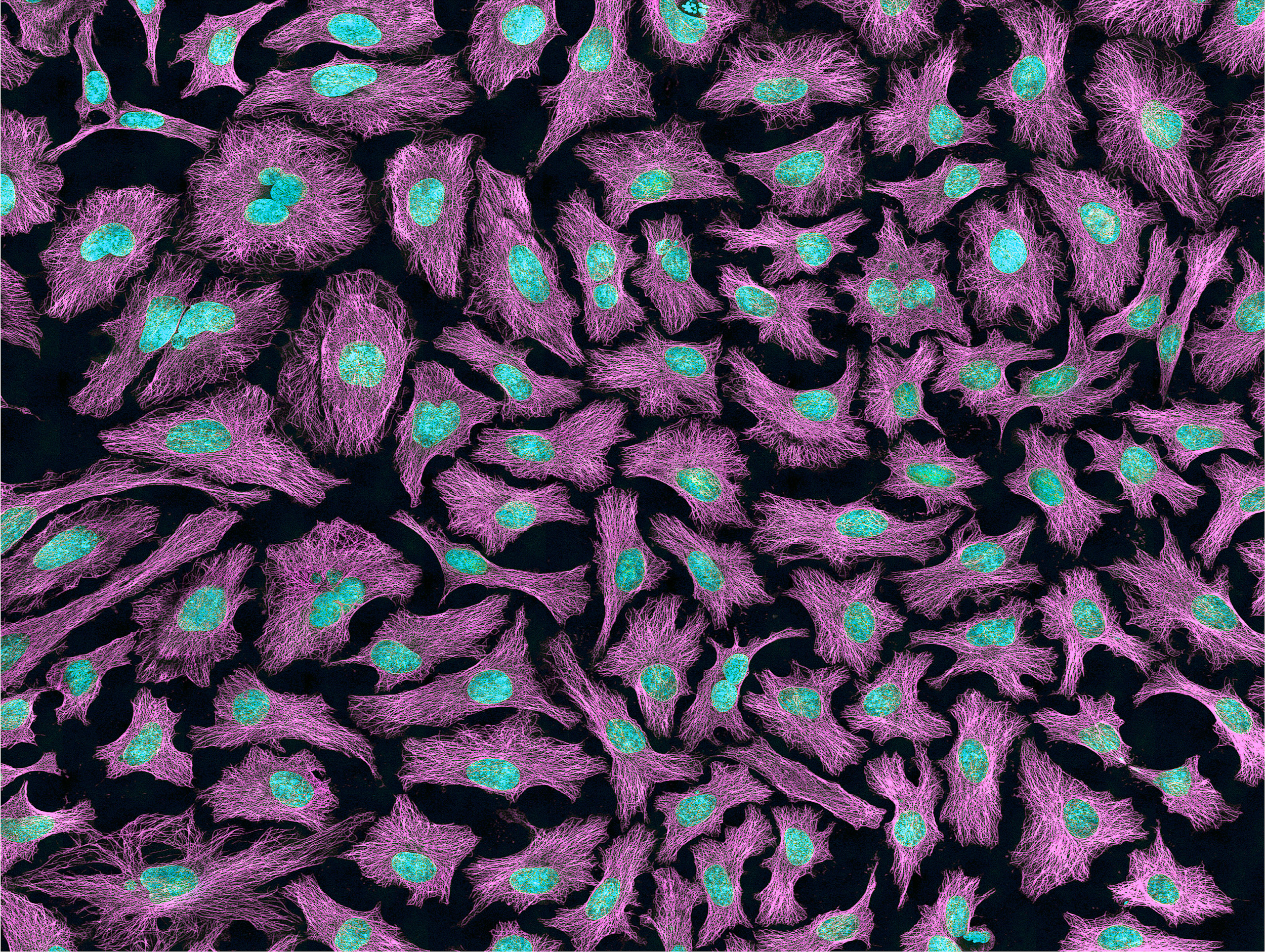 Figure \(\PageIndex{9}\): A microscopic slide of HeLa cancer cells.
Researchers have exploited the immortality of cancer cells for molecular research. The oldest immortal cell line is HeLa cells (Figure 3.19), which was harvested from Henrietta Lacks, an African-American woman diagnosed with cervical cancer in 1955. At that time, extracted cells frequently died during experiments, but surprisingly, HeLa cells continued to replicate. Propagation of Lacks’s cell line has significantly contributed to medical research, including ongoing cancer research and helping to test the polio vaccine in the 1950s. Unfortunately, Lacks had not given her consent for her tumor biopsy to be used in cell culture research. Moreover, her family was unaware of the extraction and remarkable application of her cells for two decades. The history of HeLa cell origin was first revealed in 1976. The controversy voiced by the Lacks family was included in an extensive account of HeLa cells published in Rebecca Skloot’s 2010 book, The Immortal Life of Henrietta Lacks. A film based on the book was also released in 2017.
Mitotic Cell Division
The body and its various tissues are comprised of somatic cells. Organisms that contain two sets of chromosomes in their somatic cells are called diploid organisms. Humans have 46 chromosomes and they are diploid because they inherit one set of chromosomes (n = 23) from each parent. As a result, they have 23 matching pairs of chromosomes, which are known as homologous chromosomes. These homologous pairs vary in size and are generally numbered from largest (chromosome 1) to smallest (chromosome 22), as seen in Figure 3.15, with the exception of the 23rd pair, which is made up of the sex chromosomes (X and Y). Typically, the female sex is XX and the male sex is XY. Individuals inherit an X chromosome from their mother and an X or Y from their father.
In order to grow and repair tissues, somatic cells must divide. As discussed previously, a cell must first replicate its genetic material for cell division to occur. During DNA replication, each chromosome produces double the amount of genetic information. The duplicated arms of chromosomes are known as sister chromatids, and they are attached at the centromeric region. To elaborate, the number of chromosomes stays the same (n = 46); however, the amount of genetic material is doubled in the cell as the result of replication.
Mitosis is the process of somatic cell division that gives rise to two diploid daughter cells. Figure 3.20 shows a brief overview of mitosis. Once DNA and other organelles in the cell have finished replication, mitotic spindle fibers (microtubules) assist with chromosomal movement by attaching to the centromeric region of each chromosome. Specifically, the spindle fibers physically align each chromosome at the center of the cell. Next, the spindle fibers divide the sister chromatids and move each one to opposite sides of the cell. At this phase, there are 46 chromosomes on each side of the cell. The cell can now divide into two fully separated daughter cells.
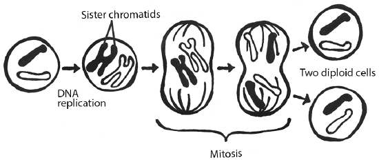 Figure \(\PageIndex{10}\): Steps of mitotic cell division.
Meiotic Cell Division
Gametogenesis is the production of gametes (sperm and egg cells); it involves two rounds of cell division called meiosis. Similar to mitosis, the parent cell in meiosis is diploid. However, meiosis has a few key differences, including the number of daughter cells produced (four cells, which require two rounds of cell division to produce) and the number of chromosomes each daughter cell has (Figure 3.21). During the first round of division (known as meiosis I), each chromosome (n = 46) replicates its DNA so that sister chromatids are formed. Next, with the help of spindle fibers, homologous chromosomes align near the center of the cell and sister chromatids physically swap genetic material. In other words, the sister chromatids of matching chromosomes cross over with each other at matching DNA nucleotide positions. The occurrence of homologous chromosomes crossing over, swapping DNA, and then rejoining segments is called genetic recombination. The “genetic shuffling” that occurs in gametes increases organismal genetic diversity by creating new combinations of genes on chromosomes that are different from the parent cell. Genetic mutations can also arise during recombination. For example, there may be an unequal swapping of genetic material that occurs between the two sister chromatids, which can result in deletions or duplications of DNA nucleotides. Once genetic recombination is complete, homologous chromosomes are separated and two daughter cells are formed.
The daughter cells after the first round of meiosis are haploid, meaning they only have one set of chromosomes (n = 23). During the second round of cell division (known as meiosis II), sister chromatids are separated and two additional haploid daughter cells are formed. Therefore, the four resulting daughter cells have one set of chromosomes (n = 23), and they also have a genetic composition that is not identical to the parent cells nor to each other.
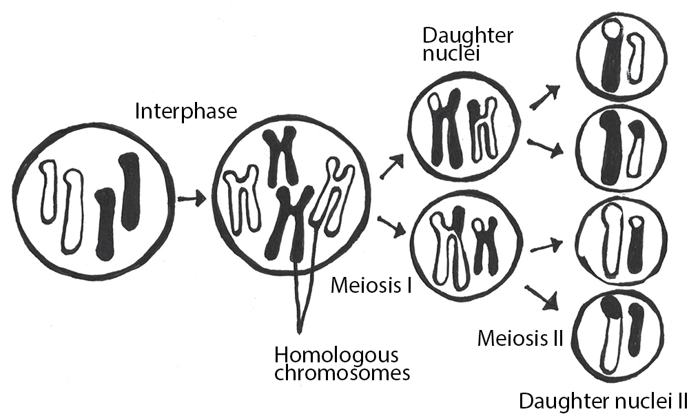 Figure \(\PageIndex{11}\): Meiotic cell division.
Although both sperm and egg gamete production undergo meiosis, they differ in the final number of viable daughter cells. In the case of spermatogenesis, four mature sperm cells are produced. Although four egg cells are also produced in oogenesis, only one of these egg cells will result in an ovum (mature egg). During fertilization, an egg cell and sperm cell fuse, which creates a diploid cell that develops into an embryo. The ovum also provides the cellular organelles necessary for embryonic cell division. This includes mitochondria, which is why humans, and most other multicellular eukaryotes, have the same mtDNA sequence as their mothers.
Chromosomal Disorders
During mitosis or meiosis, entire deletions or duplications of chromosomes can occur due to error. For example, homologous chromosomes may fail to separate properly, so one daughter cell may end up with an extra chromosome while the other daughter cell has one less. Cells with an unexpected (or abnormal) number of chromosomes are known as aneuploid. Adult or embryonic cells can be tested for chromosome number (karyotyping). Aneuploid cells are typically detrimental to a dividing cell or developing embryo, which can lead to a loss of pregnancy. However, the occurrence of individuals being born with three copies of the 21st chromosome is relatively common; this genetic condition is known as Down Syndrome. Moreover, human males and females can be born with aneuploid sex chromosome conditions such as XXY, XXX, and XO (referring to only one X chromosome).