At the beginning of the chapter, we defined proteins as strings of amino acids that fold into complex 3-D shapes. There are 20 standard amino acids that can be strung together in different orders in humans, and the result is that proteins can perform an impressive amount of different functions. For instance, muscle fibers are proteins that help facilitate movement. A special class of proteins (immunoglobulins) help protect the organism by detecting disease-causing pathogens in the body. Protein hormones, such as insulin, help regulate physiological activity. Blood hemoglobin is a protein that transports oxygen throughout the body. Enzymes are also proteins, and they are catalysts for biochemical reactions that occur in the cell (e.g., metabolism). Larger-scale protein structures can be visibly seen as physical features of an organism (e.g., hair and nails).
Transcription and Translation
Coding nucleotides in our DNA provide instructions on how to make proteins. Making proteins, also known as protein synthesis, can be broken down into two main steps referred to as transcription and translation. Protein synthesis relies on many molecules in the cell including different types of regulatory proteins and RNAs for each step in the process. Although there are many different types of RNA molecules that have a variety of functions within the cell, we will mainly focus on .
A gene is a segment of DNA that codes for RNA, and genes can vary in length from a few hundred to as many as two million base pairs in length. The purpose of transcription is to make an RNA copy of that genetic code (Figure 3.22). Unlike double-stranded DNA, RNA molecules are single-stranded nucleotide sequences (refer back to Figure 3.2). Additionally, while DNA contains the nucleotide thymine (T), RNA does not—instead, its fourth nucleotide is uracil (U). Uracil is complementary to (or can pair with) adenine (A), while cytosine (C) and guanine (G) continue to be complementary to each other. For transcription to proceed, a gene must first be turned “on” by the cell (see Special Topic: Genetic Regulation of the Lactase (LCT) Gene for a more detailed discussion of gene regulation). The double-stranded DNA is then separated, and one side of the DNA strand is used as a template where complementary RNA nucleotides are strung together. For example, if a DNA template is TACGGATGC, then the newly constructed mRNA sequence will be AUGCCUACG. Sometimes the end product needed by the cell is that transcribed RNA, but for protein synthesis constructing the RNA (specifically pre-messenger RNA, or pre-mRNA) is just the first step.
Figure \(\PageIndex{1}\): RNA polymerase catalyzing DNA transcription.
Genes contain segments called introns and exons. Exons are considered “coding” while introns are considered “noncoding”—meaning the information they contain will not be needed to construct proteins. When a gene is first transcribed into pre-mRNA, introns and exons are both included (Figure 3.23). However, once transcription is finished, introns are removed in a process called splicing. During splicing, a protein/RNA complex attaches itself to the pre-mRNA and removes introns and then connects the remaining exons, thus creating a shorter mature mRNA.
Figure \(\PageIndex{2}\): RNA processing is the modification of RNA, including the removal of introns, called splicing, between transcription and translation.
The process by which mRNA is “read” and amino acids chained together to form new proteins is called translation. During translation, mature mRNA is transported outside of the nucleus where it is bound to a ribosome (Figure 3.24). The nucleotides in the mRNA are read as triplets, which are called codons. Each codon corresponds to an amino acid, and this is the basis for building a protein. Continuing with our example from above, the mRNA sequence AUG-CCU-ACG codes for three amino acids. Using a codon table (Figure 3.25), AUG is a codon for methionine (Met), CCU is proline (Pro), and ACG is threonine (Thr). Therefore, the protein sequence is Met-Pro-Thr. Methionine is the most common “start codon” (AUG) for the initiation of protein translation in eukaryotes. As the ribosome moves along the mRNA, the growing amino acid chain exits the ribosome and folds into a protein (Figure 3.26). When the ribosome reaches a “stop” codon (UAA, UAG, or UGA), the ribosome stops adding new amino acids, detaches from the mRNA, and the protein is released. Folded proteins can then be used to complete a structural or functional task.
Figure \(\PageIndex{3}\): Translation of mRNA into an amino acid.Figure \(\PageIndex{4}\): This table can be used to identify which mRNA codons (sequence of three nucleotides) correspond with each of the 20 different amino acids. For example, if the codon is CAU, the first position is “C” and you would look in that corresponding row, the second position is “A” and you would look in that column. “U’ is the third position—narrowing the row and indicating that the CAU codon corresponds with the amino acid “histidine” (abbreviated “His”). The table also indicates the most common “start codon” (AUG) that correlates with Methionine, and the three “stop” codons (UAA, UAG, or UGA).Figure \(\PageIndex{5}\): Indicates levels of protein organization from the simple amino acid chain that is then folded and organized into more complex protein structures.
SPECIAL TOPIC: GENETIC REGULATION OF THE LACTASE (LCT) GENE
The LCT gene codes for a protein called lactase, an enzyme produced in the small intestine. It is responsible for breaking down the sugar “lactose” found in milk. Lactose intolerance occurs when not enough lactase enzyme is produced and, in turn, digestive symptoms occur. To avoid this discomfort, individuals may take lactase supplements, drink lactose-free milk, or avoid milk products altogether.
The LCT gene is a good example of how cells regulate protein synthesis. The promoter region of the LCT gene helps regulate whether it is transcribed or not transcribed (i.e., turned “on” or “off,” respectively). Lactase production is initiated when a regulatory protein known as a transcription factor binds to a site on the LCT promoter. RNA polymerases are then recruited; they read DNA and string together nucleotides to make RNA molecules (Figure 3.22). An LCT pre-mRNA is synthesized (made) in the nucleus, and further chemical modifications flank the ends of the mRNA to ensure the molecule will not be degraded in the cell.
Next, RNA processing occurs. A spliceosome complex removes the introns and connects exons to form the mature mRNA. Once the LCT mRNA is transported outside of the nucleus, it is bound to a ribosome, which is a multi-protein complex that includes. The ribosome of eukaryotes has two main subunits: the smaller bottom subunit that binds to the mRNA and the larger top subunit that contains binding sites (see Figure 3.24). Each tRNA has a nucleotide anticodon that recognizes an mRNA codon. When a tRNA binds to an mRNA codon in the ribosome, the tRNA transfers the corresponding amino acid. rRNA ensures the newly added amino acid is linked in the correct order. The growing protein then folds into the lactase enzyme, which can break down lactose.
Most animals lose their ability to digest milk as they mature due to the decreasing transcriptional “silence” of the LCT gene over time. However, some humans have the ability to digest lactose into adulthood (also known as “lactase persistence”). This means they have a genetic mutation that leads to continuous transcriptional activity of LCT. Lactase persistence mutations are common in populations with a long history of pastoral farming, such as northern European and North African populations. It is believed that lactase persistence evolved because the ability to digest milk was nutritionally beneficial. More information about lactase persistence will be covered in Chapter 14.

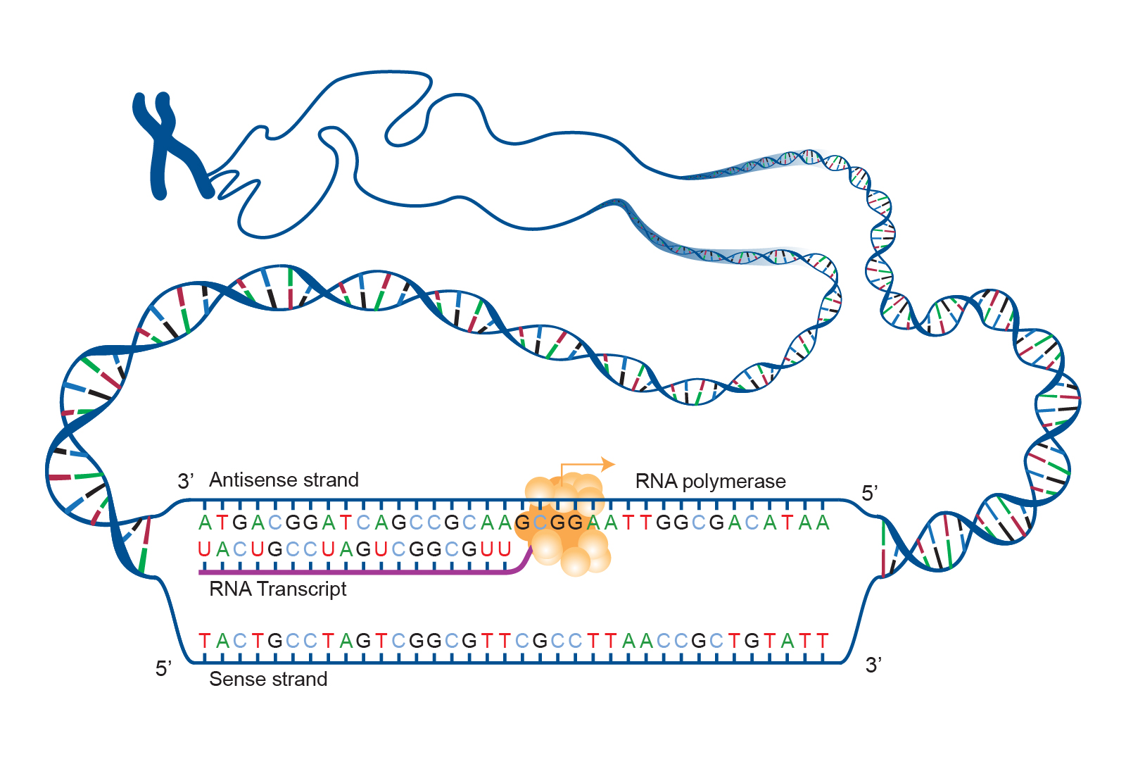 Figure \(\PageIndex{1}\): RNA polymerase catalyzing DNA transcription.
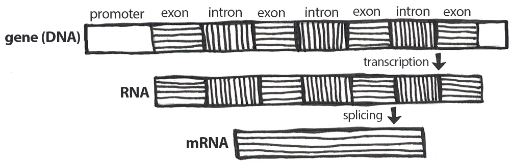 Figure \(\PageIndex{2}\): RNA processing is the modification of RNA, including the removal of introns, called splicing, between transcription and translation.
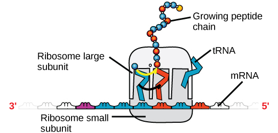 Figure \(\PageIndex{3}\): Translation of mRNA into an amino acid.
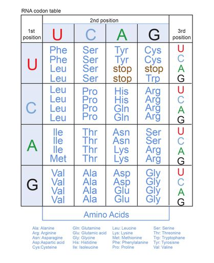 Figure \(\PageIndex{4}\): This table can be used to identify which mRNA codons (sequence of three nucleotides) correspond with each of the 20 different amino acids. For example, if the codon is CAU, the first position is “C” and you would look in that corresponding row, the second position is “A” and you would look in that column. “U’ is the third position—narrowing the row and indicating that the CAU codon corresponds with the amino acid “histidine” (abbreviated “His”). The table also indicates the most common “start codon” (AUG) that correlates with Methionine, and the three “stop” codons (UAA, UAG, or UGA).
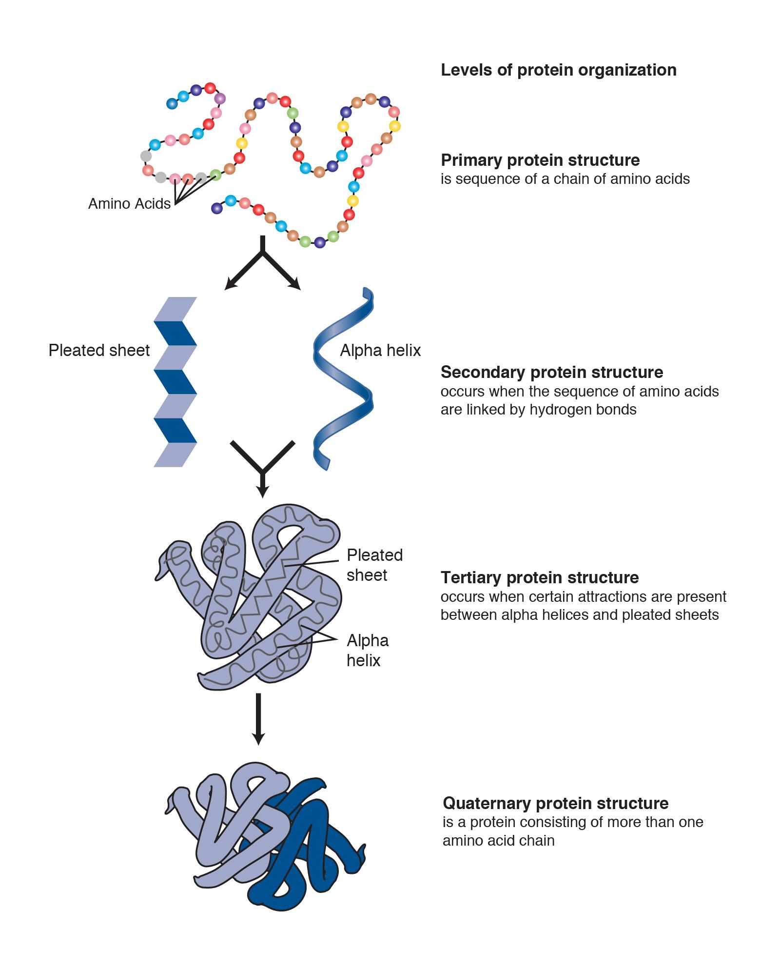 Figure \(\PageIndex{5}\): Indicates levels of protein organization from the simple amino acid chain that is then folded and organized into more complex protein structures.