Scientists have adopted standardized terminology to describe the position of the body as well as the location and movements of different body parts relative to one another. The terminology used throughout this appendix is consistent with the most recent edition of Terminologia Anatomica: International Anatomical Terminology (Federative Committee on Anatomical Terminology 1998). Below you will find descriptions of standard anatomical position, directional terms, anatomical planes, and skeletal movements (i.e., movements possible where two bones articulate through a joint).
When a body is in anatomical position, it is situated as if the individual is standing upright; with head, eyes, and feet pointing forward (anteriorly, see below); and with arms at the side and palms facing forward. In anatomical position, the bones of the forearm are not crossed Figure \(\PageIndex{1}\).
Figure \(\PageIndex{1}\): The human body is shown in anatomical position in an (left) anterior view and a (right) posterior view.
With the body in anatomical position, the position of specific organs (e.g., bones) can be described as situated within specific anatomical planes (Figure \(\PageIndex{2}\)). These imaginary planes bisect the body into equal or subequal halves, depending on which plane is described. Coronal (frontal) planes divide the body vertically into anterior (front) and posterior (back) halves. Transverse planes divide the body horizontally into superior (upper) and inferior (lower) halves. Sagittal planes divide the body vertically into left and right halves. The plane that divides the body vertically into equal left and right halves is called the midsagittal plane. The midsagittal plane is also called the median plane because it is in the midline of the body. If the left and right halves of the body are divided unequally (i.e., the right “half” is larger than the left “half” or vice versa), we call that dividing plane a parasagittal plane. There are potentially an infinite number of parasagittal planes that can be drawn to divide the body into unequal right and left “halves.”
Figure \(\PageIndex{2}\): The three planes most commonly used in anatomical and medical imaging are the sagittal, frontal (or coronal), and transverse planes.
Directional Terms
When describing the position of one body part (in this case, a bone) relative to another, scientists use precisely defined directional terms. Each of the directional terms described below refers to the body in anatomical position. This is an important point because once the position of one bone is established relative to another, that directional term is the same, regardless of whether the body remains in anatomical position (e.g., the skull is always superior to the vertebrae, even if the individual is lying down).
A bone or skeletal feature that is anterior (or ventral) is located toward the front of the body and a bone that is posterior (or dorsal) is located toward the back of the body (Figure \(\PageIndex{3}\)). For example, the sternum (breastbone) is anterior to the vertebral column (“backbone”). A medial bone is located closer to the midline (midsagittal plane) than a bone that is lateral, or located further from the midline. For example, the thumb is lateral to the index finger. A structure that is proximal is closer to the trunk of the body (usually referring to limb bones) than a distal structure, which is further from the trunk of the body. For example, the femur (thigh bone) is proximal to the tibia (leg bone). Finally, structures that are superior (or cranial) are located closer to the head than structures that are inferior (or caudal). For example, the rib cage is superior to the pelvis, and the foot is inferior to the knee. Typically, the terms “cranial” and “caudal” are used in reference to the non-human, quadrupedal skeleton, whereas “superior” and “inferior” are used in reference to the human skeleton.
Figure \(\PageIndex{3}\): Paired directional terms are shown as applied to the human body.

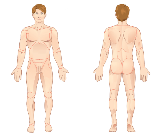 Figure \(\PageIndex{1}\): The human body is shown in anatomical position in an (left) anterior view and a (right) posterior view.
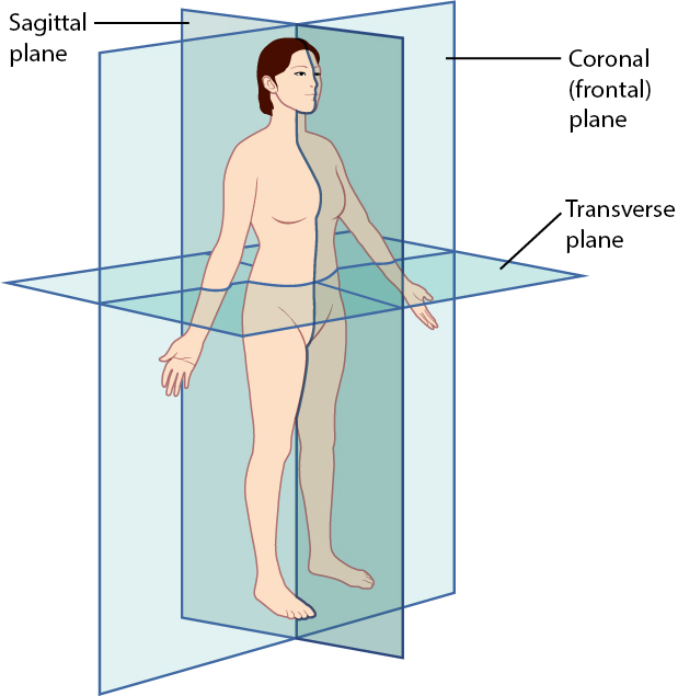 Figure \(\PageIndex{2}\): The three planes most commonly used in anatomical and medical imaging are the sagittal, frontal (or coronal), and transverse planes.
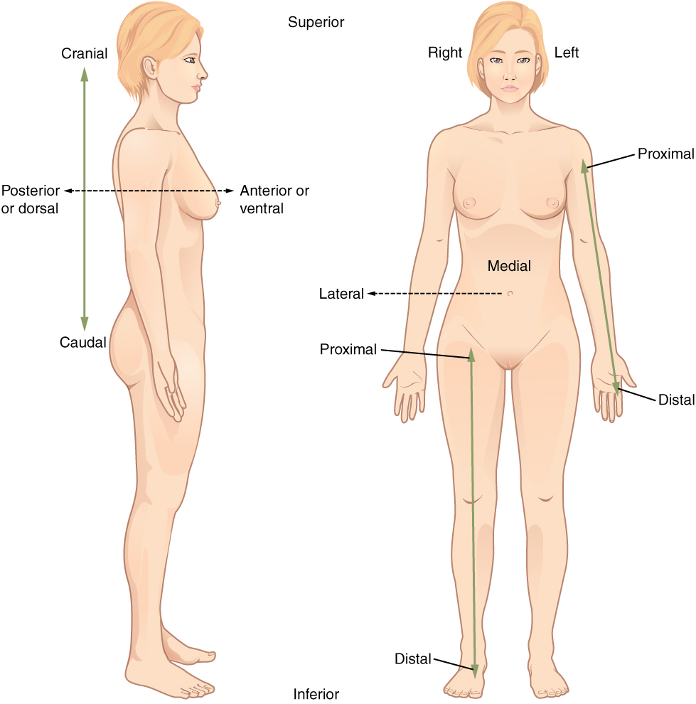 Figure \(\PageIndex{3}\): Paired directional terms are shown as applied to the human body.