Figure \(\PageIndex{1}\): A typical long bone shows the gross anatomical characteristics of bone.
Bone is a composite of organic (collagen) and inorganic (mineral, e.g., hydroxyapatite, a calcium phosphate salt) materials with incredible strength in compression so it can support the body under the force of gravity. When bone is mature (fully mineralized as opposed to juvenile and undermineralized), it comprises an outer dense region of bone called cortical (or compact) bone and an inner spongy region of bone called cancellous (or trabecular) bone (Figure A.4). However, the interfaces between the organic and inorganic materials, as well as the cortical and cancellous regions, are subject to changing stresses. Each time we move our muscles, our bones are subjected to a combination of bending, twisting, compression, and tension. This results in the formation of microscopic cracks that weaken the bone and may result in complete bone fracture. Bone cells called osteocytes have special properties that allow them to sense when these microcracks form. Osteocytes then signal osteoclast cells to remove the cracked bone and osteoblast cells to lay down new bone—a process known as skeletal remodeling. Osteogenic cells are stem cells that are able to differentiate into osteoblasts (Figure A.5).
Figure \(\PageIndex{2}\) Four types of cells are found within bone tissue. Osteogenic cells are stem cells that develop into osteoblasts. Osteoblasts lay down new bone while osteoclasts remove bone. Osteoblasts that get trapped in calcified matrix become osteocytes.
Bone Shape
Different bones have different shapes that largely relate to their specific function within the skeletal system. Additionally, the ratio of cortical to cancellous bone, and which muscles are attached to the bone and how, affect the shape of the whole bone. Generally there are five recognized bone shapes: long bones, short bones, flat bones, sesamoid bones, and irregular bones. Long bones are longer than they are wide and consist of three sections: diaphysis, epiphysis, and metaphysis (Figure A.4). The diaphysis of a long bone is simply the shaft of the bone, and it comprises mostly cortical bone with a thin veneer of internal cancellous bone lining a medullary cavity. At both the proximal and distal ends of every long bone, there is an epiphysis, which consists of a thin shell of cortical bone surrounding a high concentration of cancellous bone. The epiphysis is usually coated with hyaline (or articular) cartilage to facilitate joint articulation with other bones. The junction between diaphysis and epiphysis is the metaphysis, which has a more equal ratio of cortical to cancellous bone. Examples of long bones are the humerus, the femur, and the metacarpals and metatarsals.
The other bone shapes are simpler. Short bones are defined as being equal in length and width, and they possess a mix of cortical and cancellous bone (Figure A.6). They are usually involved in forming movable joints with adjacent bones and therefore often have surfaces covered with hyaline cartilage. Examples of short bones are the carpals of the wrist and the tarsals of the ankle.
Flat bones, as their name suggests, are flat and consist of two layers of thick cortical bone with an intermediate layer of cancellous bone called a diploe. Examples of flat bones are most of the bones of the skull, such as the frontal and parietal bones, as well as all parts of the sternum (Figure A.6). Sometimes bones develop within the tendon of a muscle in order to reduce friction on the joint surface and to increase leverage of the muscle to move a joint. These types of bones are called sesamoid bones, and these include the patella (or knee cap) and the pisiform (a bone of the wrist).
Irregular bones are bones that don’t fit into any of the other four categories. The shapes of these bones are often more complex than the others, and examples include the vertebrae and certain bones of the skull, like the ethmoid and sphenoid bones (Figure A.6).
Figure \(\PageIndex{3}\) Bones are classified according to their shape and include long, short, flat, sesamoid, and irregular bones.
Bone Formation
Bone develops via one of two mechanisms: intramembranous or endochondral bone formation. Intramembranous bone formation occurs when connective tissue mesenchymal (stem) cells aggregate and differentiate into osteoblasts, which then begin to synthesize new bone along the aggregated connective tissue cells (Figure A.7). Intramembranous bone formation is the mechanism by which most bones of the skull develop as well as the clavicle (collar bone). When osteoblasts develop from an intermediate cartilage “model” that is then replaced by bone, instead of developing directly from the mesenchymal cells, the mechanism is described as endochondral bone formation (Figure A.8). Endochondral bone formation is the mechanism by which most bones of the skeleton develop (Burr and Organ 2017).
Figure \(\PageIndex{4}\) Intramembranous ossification begins when mesenchymal cells group into clusters. These clusters contain osteoblasts, which lay down the initial trabecular bone. Compact bone develop superficial to the trabecular bone, and the initial structure of the bone is complete.Figure \(\PageIndex{5}\) Endochondral ossification begins when mesenchymal cells differentiate into cartilage cells which lay down a cartilage model of the future bony skeleton. Cartilage is then replaced by bone, except at the (epiphyseal) growth plates (which fuse at the end of postnatal growth) and the hyaline (articular) cartilage on the joint surface.
Bone Function
Bone performs both metabolic and mechanical functions for the body. On the metabolic side, bone is required to maintain mineral (i.e., calcium) homeostasis and for the production of red and white blood cells (Figure A.9), which develop in the cavity and the cancellous region of the metaphysis and epiphysis. But it is undeniable that the mechanical functions of bone are primary because bone is critically responsible for protecting internal organs, providing support against the force gravity, and serving as a network of rigid levers for muscles to act upon during movement.
Figure \(\PageIndex{6}\) The body regulates calcium homeostasis with two pathways; one is signaled to turn on when blood calcium levels drop below normal and one is the pathway that is signaled to turn on when blood calcium levels are elevated.

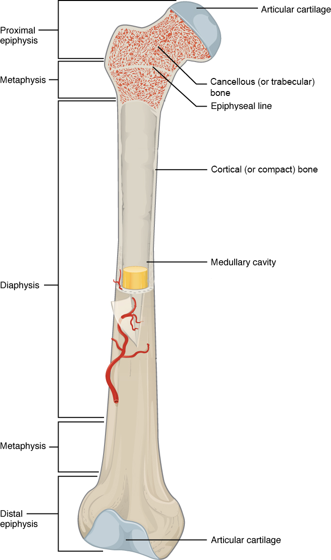 Figure \(\PageIndex{1}\): A typical long bone shows the gross anatomical characteristics of bone.
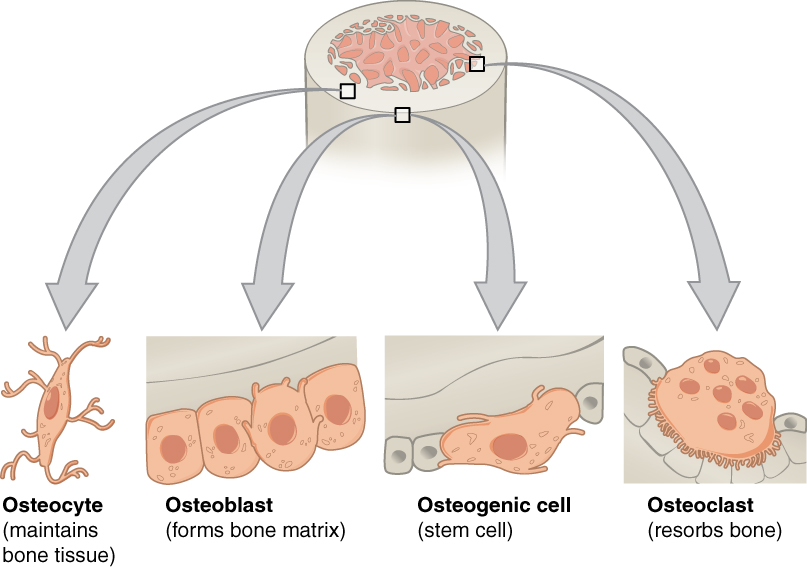 Figure \(\PageIndex{2}\) Four types of cells are found within bone tissue. Osteogenic cells are stem cells that develop into osteoblasts. Osteoblasts lay down new bone while osteoclasts remove bone. Osteoblasts that get trapped in calcified matrix become osteocytes.
 Figure \(\PageIndex{3}\) Bones are classified according to their shape and include long, short, flat, sesamoid, and irregular bones.
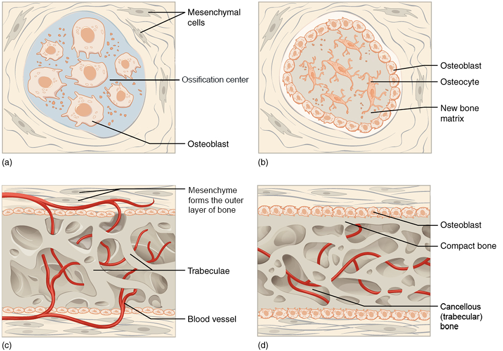 Figure \(\PageIndex{4}\) Intramembranous ossification begins when mesenchymal cells group into clusters. These clusters contain osteoblasts, which lay down the initial trabecular bone. Compact bone develop superficial to the trabecular bone, and the initial structure of the bone is complete.
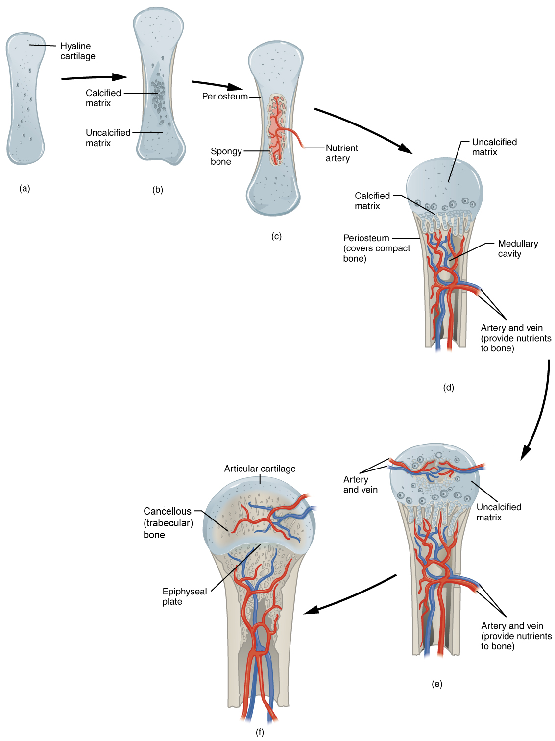 Figure \(\PageIndex{5}\) Endochondral ossification begins when mesenchymal cells differentiate into cartilage cells which lay down a cartilage model of the future bony skeleton. Cartilage is then replaced by bone, except at the (epiphyseal) growth plates (which fuse at the end of postnatal growth) and the hyaline (articular) cartilage on the joint surface.
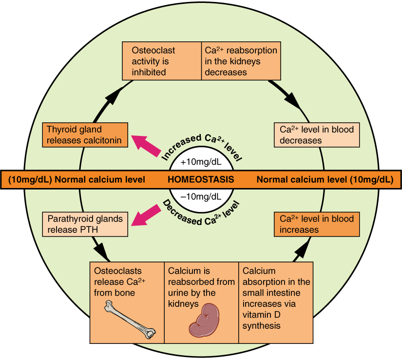 Figure \(\PageIndex{6}\) The body regulates calcium homeostasis with two pathways; one is signaled to turn on when blood calcium levels drop below normal and one is the pathway that is signaled to turn on when blood calcium levels are elevated.