The skeletal system is divided into two regions: axial and appendicular (Figure A.10). The axial skeleton consists of the skull, vertebral column, and the thoracic cage formed by the ribs and sternum (breastbone). The appendicular skeleton comprises the pectoral girdle, the pelvic girdle, and all the bones of the upper and lower limbs (White and Folkens 2000).
Figure \(\PageIndex{1}\): The axial skeleton consists of the skull, vertebral column, and the thoracic cage. The appendicular skeleton is made up of all bones of the upper and lower limbs.
Axial Skeleton
Skull
The skull comprises numerous bones (some paired and others that are unpaired) and is divided into two major portions: the mandible (or lower jaw) and the cranium (the remainder of the skull). The cranium is further subdivided into the neurocranium (or cranial vault), which houses the brain, and the viscerocranium (or facial skeleton; Figure A.11). Where two bones of the cranium come together, they form articulations called cranial sutures, which fuse (or close) with increasing age and can be used as a broad estimate of age at death. Degree of suture closure is scored at several anatomical landmarks and compiled to produce an age estimate. The remainder of this section lists the bones of the skull by region and details some of the landmarks examined in forensic contexts.
Figure \(\PageIndex{2}\): The skull consists of the cranium and the mandible. The cranium is further divided into the neurocranium and viscerocranium.
Bones and Some Features of the Neurocranium
Frontal: an unpaired bone consisting of two parts: a superior, vertically oriented portion called the squama and an inferior, horizontally oriented portion that forms the roof of the orbit (eye socket; Figures A.12 and A.13).
The coronal suture is the articulation between the frontal bone and the two parietal bones posterior and lateral to the frontal.
The frontal bone develops initially as two separate bones that fuse together during growth. Occasionally this fusion is incomplete, resulting in a metopic suture that persists between the two halves (left and right) of the frontal bone (White and Folkens 2005).
The glabella is a bony projection between the brow ridges. The glabella in females tends to be flat while rounded and protruding in males.
The supraorbital margin is the upper edge of the orbit. The thickness of the edge can be used as an indicator of sex. A thin, sharp border is indicative of a female while a blunt, thick border suggests a male.
Figure \(\PageIndex{3}\): Anterior view of the skull.Figure \(\PageIndex{4}\): Lateral view of the skull.
Parietal: Paired bones that form the majority of the roof and sides of the neurocranium (Figures A.12 and A.13).
The sagittal suture is the articulation between the right and left parietal bones. It extends from the coronal suture anteriorly to the lambdoidal suture, which separates the parietal bones from the occipital bone posteriorly.
Each parietal bone is marked by two temporal lines (superior and inferior), which are anterior-posterior arching lines that serve as attachment sites for a major chewing muscle (temporalis) and its associated connective tissue.
Temporal: Paired bones on the lateral side of the neurocranium that are divided into two portions: squamous (or flat) portion that forms the lateral side of the neurocranium and the petrous (or rock-like) portion that houses the special sense organs of the ear for hearing and balance as well as the three tiny bones of the middle ear: incus, malleus, and stapes (Figures A.13, A.14, and A.15).
The squamosal suture is the articulation between the squamous portion of the temporal bone and the inferior border of the parietal bone.
The mastoid process is a prominent attachment site for several muscles including the large sternocleidomastoid muscle. As such, it is often used to estimate sex in that males tend to have longer and wider mastoid processes compared to females (Bass 2005).
The styloid process is a thin, pointed, inferior projection of the temporal bone that serves as an attachment site for several muscles and a ligament of the throat.
The zygomatic processof the temporal is a thin, arch-like process that originates from the squamous portion of the temporal bone. The zygomatic process articulates with the temporal process of the zygomatic bone to form the zygomatic arch (or cheekbone).
The temporal fossa is the depression in the temporal bone where the mandibular condyle (see below, under mandible) articulates to form the temporomandibular (or jaw) joint.
Figure \(\PageIndex{5}\): A lateral view of the isolated temporal bone shows the squamous portion.Figure \(\PageIndex{6}\): (a) The base of the cranium (b) The floor of the cranial cavity.
Occipital: Unpaired bone that forms the posterior and inferior portions of the neurocranium (Figures A.13 and A.15).
The lambdoidal suture is the articulation between the occipital bone and the two parietal bones. It resembles the shape of the Greek letter lambda.
The external occipital protuberance (EOP) is a robust attachment of the nuchal ligament. When viewed laterally, males tend to have a discernible projection or hook, whereas in females, the occipital is typically smooth.
The nuchal lines are parallel ridges that meet on the midline at the EOP. These prominent muscle attachment sites tend to be more robust and projecting in males.
The occipital bone contains a large circular opening called the foramen magnum, which provides a space for passage of the brainstem/spinal cord from the neurocranium into the vertebral canal of the spine.
Sphenoid: Unpaired, butterfly-shaped bone that forms the central portion of the bottom of the neurocranium. The sphenoid is divided into several regions, including the body, greater wings, lesser wings, and pterygoid processes (with pterygoid plates; see Figures A.15 and A.16). This bone is critical to supporting the brain and several nerves and blood vessels supplying this region.
Pterygoid plates are flat projections of the pterygoid processes that serve as attachment sites for chewing muscles and muscles of the throat.
Figure \(\PageIndex{7}\): Shown in isolation in (a) superior and (b) posterior views, the sphenoid forms the central portion of the neurocranium. The sphenoid has multiple openings for the passage of nerves and blood vessels.
Ethmoid: Unpaired bone consisting of a median vertical plate that forms part of the bony nasal septum and a horizontal plate (cribriform plate) with many small foramina (holes) that transmit olfactory nerves (special sense of smell; Figure A.17).
Figure \(\PageIndex{8}\): The unpaired ethmoid bone is located at the midline within the central skull. It forms the upper nasal septum and contains foramina to convey olfactory nerves.
Bones of the Viscerocranium
Maxilla: Paired bones that form the upper jaw, support the upper teeth, and form the inferior margin of the cheek (Figures A.12, A.13, A.15, and A.18).
The nasal spine is a thin projection on the midline at the inferior border of the nasal aperture. Length of the nasal spine has been used as a subjective trait to determine ancestry, with those exhibiting long nasal spines to be more likely of European ancestry.
The zygomatic process of the maxilla is the portion of the bone that articulates with the zygomatic bone to form the anterior portion of the zygomatic arch.
Figure \(\PageIndex{9}\): The maxilla forms the upper jaw and supports the upper teeth.
Nasal: Small, paired, flat, rectangular bones that form the bridge of the nose (Figure A.19).
Nasal aperture is the anterior opening into the nasal cavity. As a forensic trait, it is described as low and wide in those of African ancestry and tall and narrow in those of European ancestry.
Zygomatic: Paired bones that form the anterolateral portion of the cheekbone and contribute to the lateral and inferior wall of the orbit (Figure A.19).
The temporal process of the zygomatic is the portion of the bone that articulates with the temporal bone to form the anterior portion of the zygomatic arch.
Palatine: Paired L-shaped bones that form the posterior portion of the roof of the mouth, floor of the orbit, and the floor and lateral walls of the nasal cavity (Figures A.15 and A.19).
Lacrimal: Small, flat, paired bones that form the anterior portion of the medial wall of the orbit (Figure A.19).
Figure \(\PageIndex{10}\): Seven skull bones contribute to the walls of the orbit: frontal, zygomatic, maxilla, lacrimal, ethmoid, palatine, and sphenoid.Figure \(\PageIndex{11}\): The nasal septum is formed by the perpendicular plate of the ethmoid bone and the vomer bone.
Vomer: Unpaired thin bone that forms the inferior portion of the bony nasal septum. It articulates with the ethmoid superiorly (Figure A.20).
Inferior nasal concha: Paired bones that project and curl like a scroll from the lateral wall of the nasal cavity (Figure A.21).
Figure \(\PageIndex{12}\): Inferior nasal concha scroll from the lateral wall of the nasal cavity.
Hyoid: Unpaired U-shaped bone that sits in the neck inferior to the mandible. The hyoid is the only bone of the skeleton that does not articulate with another bone. Instead, it is encased in a sling of muscles that move the larynx (voice box), pharynx, and tongue (Figure A.22).
Figure \(\PageIndex{13}\): The hyoid bone is located in the upper neck and does not join with any other bone. It provides attachments for muscles that move the tongue, larynx, and pharynx.
Mandible: Unpaired bone with a horizontal (and anteriorly arched) body and a vertical ramus that articulates with the temporal fossa to form the temporomandibular (jaw) joint. The body of the mandible houses the lower teeth (Figure A.13 and A.23).
The mental protuberance (eminence) is the most anteriorly projecting point on the mandible—the so-called “chin.” A prominent projection is likely to indicate a male, while a smooth mental region likely indicates a female.
The ramus of the mandible projects superiorly from the body of the mandible and ascends to one of two features on the superior aspect: coronoid process or mandibular condyle.
The coronoid process is a bony projection off the anterior and superior aspect of the mandibular ramus. The inferior attachment of the temporalis muscle (a chewing muscle) attaches here.
The mandibular condyle, a rounded projection off the posterior and superior aspect of the mandibular ramus, articulates with the temporal fossa of the temporal bone at the temporomandibular (TMJ) joint.
The gonial (mandibular) angle is the rounded posteroinferior border of the mandible. It tends to be smooth in females with a more obtuse angle but is laterally flared in males and closer to a right angle in shape.
Figure \(\PageIndex{14}\): The mandible articulates with the temporal fossa to form the temporomandibular joint.
Teeth: Adults normally have 32 teeth, distributed among four quadrants of the mouth (upper left, upper right, lower left, lower right). In each quadrant, there are eight teeth: two incisors (central and lateral), one canine, two premolars, and three molars. Each of these types of teeth has a different shape that reflects its function during chewing:
Incisors are flat and shovel shaped and are used to bite into a food item.
Canines are conical, with a single pointed cusp used to puncture a food item.
Premolars have two rounded cusps and are used to grind and mash a food item.
Molars have five flatter cusps and are used to grind food prior to swallowing.
The teeth have their own set of directional terms that help differentiate the different parts of the tooth. For example, the anterior portion of the tooth is called mesial, while the posterior portion of the tooth is called distal. In the case of teeth in the front of the mouth, mesial refers to the aspect toward the midline of the body; distal refers to the aspect away from the midline. Similarly, the side of the tooth facing the lips is called the buccal surface and the side facing the tongue is called the lingual surface. Finally, we can talk about the occlusal surface of the tooth, which is the surface that comes in contact with food or the teeth from the other jaw when the jaw is closed. Sometimes the occlusal surface of the incisors is called the incisal surface.
Vertebral Column
The adult vertebral column consists of 32–33 individual vertebrae, divided into five regions: cervical, thoracic, lumbar, sacral, and coccygeal. This section will review the features of a general vertebra and then will describe the unique features of vertebrae in each of the five regions.
General Structure of a Vertebra
A typical vertebra consists of an anteriorly situated body—the main weight-bearing element of the vertebra—and a posteriorly projecting vertebral arch (Figure A.24). The vertebral arch consists of the paired pedicles and paired laminae. The pedicle connects the transverse process (a laterally projecting process that serves as an attachment site for muscles and ligaments) to the vertebral body; the lamina connects the spinous process (a posteriorly projecting process that serves as an attachment site for muscles and ligaments) to the transverse process. Projecting inferiorly off the vertebral arch is the inferior articular process, and projecting superiorly off the vertebral arch is the superior articularprocess. Between the vertebral body anteriorly and the vertebral arch posteriorly is an open space called the vertebral foramen.
Adjacent vertebrae articulate with one another through two major types of joints: intervertebral disc joints between adjacent vertebral bodies and zygapophyseal (facet) joints between the inferior articular process of one vertebra and the superior articular process of the vertebra immediately inferior to it. When all vertebrae are articulated into a column, the adjacent vertebral foramina form the vertebral canal, through which the spinal cord travels from the foramen magnum of the occipital bone to approximately the level of the second lumbar vertebra. At the level of each vertebra, the spinal cord gives us a pair (left and right) of spinal nerves that exit between adjacent vertebrae through the intervertebral foramen formed by adjacent vertebral arches. Even though the spinal cord ends in the lumbar region, the spinal nerves emanating from the spinal cord continue all the way to the sacrum (sometimes to the coccyx), culminating in a total of 30–31 pairs of spinal nerves.
Figure \(\PageIndex{15}\): A typical vertebra consists of a body and a vertebral arch. The arch is formed by the paired pedicles and paired laminae. Arising from the vertebral arch are the transverse, spinous, superior articular, and inferior articular processes. The vertebral foramen provides for passage of the spinal cord. Each spinal nerve exits through an intervertebral foramen, located between adjacent vertebrae.
Regional Differences in Vertebral Shape
In the cervical region of the vertebral column, there are seven vertebrae (named C1–C7 from superior to inferior; Figure A.25). The first two cervical vertebrae are unique from each other and all other cervical vertebrae, and they get special names: atlas (C1) and axis (C2). The atlas lacks a vertebral body (having only two large articular facets for articulation with the occipital bone of the skull: the atlanto-occipital joint for nodding the head) and does not have a spinous process. The axis is notable for the superiorly projecting dens (or odontoid process), which articulates with the atlas to create the atlanto-axial joint for head rotation. Otherwise, a typical cervical vertebra has a small vertebral body, a bifid (split) spinous process, a transverse process with a transverse foramen on it for passage of the vertebral artery and vein, and a triangular-shaped vertebral foramen.
Figure \(\PageIndex{16}\): A typical cervical vertebra has a small body, a bifid spinous process, transverse processes that have a transverse foramen, and a triangular vertebral foramen. The atlas (C1 vertebra) does not have a body or spinous process. The axis (C2 vertebra) has the upward projecting dens, which articulates with the atlas.
The vertebrae in the other regions of the spinal column are less variable in shape than the cervical region vertebrae. There are 12 thoracic region vertebrae (T1–T12), and they can be easily distinguished from the vertebrae in other regions because they have articular facets on their vertebral bodies for articulation with the head of a rib, as well as articular facets on the transverse process for articulation with the rib tubercle (Figure A.26). In particular, the vertebral bodies of T2–T9 have two pairs of articular facets called demifacets (superior and inferior), for articulation with multiple ribs; T1 and T10–T12 have single facets for articulation with a single rib. All five lumbar region vertebrae (L1–L5) are distinguished by their large vertebral body and rounded spinous process (Figure A.27). Finally, there is the sacrum, which is a bone of the pelvis that forms from the fusion of all five sacral region vertebrae (S1–S5), and there is the coccyx, which comprises three to four fused coccygeal region vertebrae that form the tailbone (Figure A.28). All great apes (including humans) lack an external tail; the coccygeal vertebrae are homologs of the external tail vertebrae in other primates and mammals (Organ 2017). Homologs are anatomical features that have the same evolutionary origin but do not necessarily have identical structure or function (i.e., the wings of bats and the arms of humans are homologous).
Figure \(\PageIndex{17}\): Thoracic vertebrae have superior and inferior articular facets on the vertebral body for articulation with the head of a rib, and a transverse process facet for articulation with the rib tubercle.Figure \(\PageIndex{18}\): Lumbar vertebrae are characterized by having a large, thick body and a short, rounded spinous process.Figure \(\PageIndex{19}\): The sacrum is formed from the fusion of five sacral vertebrae, whose lines of fusion are indicated by the transverse ridges. The coccyx is formed by the fusion of three to four coccygeal vertebrae.
Curvatures of the Vertebral Column
The adult spine is curved in the midsagittal plane in four regions of the vertebral column (cervical, thoracic, lumbar, and sacral; Figure A.29). During the fetal period of development, the vertebral column develops an anteriorly concave curvature called a kyphosis. But during the postnatal period, when an infant learns to hold its head up and then again when it learns to walk, it develops secondary curvatures called lordoses (singular: lordosis) that are posteriorly concave in the cervical and lumbar vertebral regions, while the kyphoses remain in the thoracic and sacral regions. The end result is an S-shaped curvature to our spine that enables us to keep our head and torso above our center of mass (near our pelvis) while walking around on two legs.
Figure \(\PageIndex{20}\): The adult vertebral column is curved in the midsagittal plane, with two primary curvatures (thoracic and sacral kyphoses) and two secondary curvatures (cervical and lumbar lordoses).
Thoracic Cage
The thoracic cage is formed from the sternum and the 12 ribs and their cartilages (costal cartilages), and the 12 thoracic vertebrae with which the ribs articulate (Figure A.30). The sternum comprises the manubrium (superior portion), the body of the sternum, and the xiphoid process. Each rib has a head and neck (with rib tubercle) at the vertebral end of the rib as well as a flattened shaft that extends to articulate with the sternum. All ribs articulate with the vertebral column at two points: the transverse process facet (rib tubercle) and vertebral body articular facet (head of rib). But articulations between the ribs and the sternum vary, where some ribs (1–7, the “true ribs”) attach directly to the sternum via their costal cartilages, other ribs (8–10, the “false ribs”) attach indirectly to the sternum via the costal cartilage of the rib above, and some ribs (11–12, the “floating ribs”) do not attach to the sternum at all. With increasing age, the sternal end of the rib becomes thinner and irregularly shaped compared to the smooth, rounded shape seen in young adults.
Figure \(\PageIndex{21}\): The thoracic cage is formed by the (a) sternum and (b) 12 pairs of ribs with their costal cartilages. The ribs are anchored posteriorly to the 12 thoracic vertebrae. The sternum consists of the manubrium, body, and xiphoid process. The ribs are classified as true ribs (1–7) and false ribs (8–12). The last two pairs of false ribs are also known as floating ribs (11–12).
Appendicular Skeleton
Pectoral Girdle
The pectoral girdle consists of the clavicle and the scapula, and it serves as the proximal base of the upper limb as well as the anchor for the upper limb to the axial skeleton. The clavicle is an S-shaped bone, and it forms the strut that connects the scapula to the sternum (Figure A.31). The scapula is a large, flat bone with three angles (superior, inferior, and lateral) and three borders (medial, lateral, and superior). The lateral angle is noteworthy because it serves as the articulation for the head of the humerus of the upper limb at the glenoid cavity (or glenoidfossa; Figure A.32). The borders and the anterior and posterior surfaces of the scapula are sites of muscle attachment. The scapula also has three important projections for muscle and ligament attachments: the coracoid process anteriorly and superiorly; the acromion, which articulates with the lateral end of the clavicle; and the spine on the posterior aspect of the scapula.
Figure \(\PageIndex{22}\): The scapula is shown from its anterior (deep) side and its posterior (superficial) side.
Upper Limb
The bones of the upper limb skeleton include the humerus, radius, ulna, eight carpal (wrist) bones, five metacarpal (hand) bones, and 14 phalanges (finger bones). Each of these bones is described below along with several of the prominent features.
The humerus is the bone of the arm. On the proximal epiphysis of the humerus are attachment sites for muscles of the rotator cuff (greater tubercle and lesser tubercle). A major shoulder muscle (deltoid muscle) attaches to the humerus along the lateral aspect of the diaphysis at the deltoidtuberosity. On the distal epiphysis of the humerus, the medial epicondyle is an attachment site for muscles that flex the forearm, and the lateral epicondyle is an attachment site for muscles that extend the forearm (Figure A.33).
Figure \(\PageIndex{23}\): The humerus is the single bone of the upper arm region. It articulates with the radius and ulna bones of the forearm to form the elbow joint.
There are two bones of the forearm, attached to each other by a thick connective tissue interosseous membrane: the radius and the ulna (Figure A.34). The radius is lateral to the ulna in anatomical position (this is called supination of the forearm), but it crosses over the ulna when the wrist is rotated so that the thumb points medially (this is called pronation of the forearm). On the proximal end of the radius is the radial tuberosity, an attachment site for the biceps brachii muscle that will help supinate and flex the forearm; on the distal end of the radius is the styloid process, an attachment site for ligaments of the wrist. The ulna also has a styloid process, but unlike the one on the radius it does not have a relevant function. Instead, the important processes on the ulna are located proximally, and they include the olecranonprocess for the attachment of the triceps brachii muscle (a muscle that extends the forearm and arm) and the coronoid process for the attachment of the brachialis muscle (a muscle that flexes the forearm).
Figure \(\PageIndex{24}\): The ulna is located on the medial side of the forearm, and the radius is on the lateral side. These bones are attached to each other by an interosseous membrane.
There are eight carpal bones that comprise the wrist, and they are organized into two rows: proximal and distal (Figure A.35). The proximal row of carpals (from lateral to medial) includes the scaphoid, lunate, triquetrum, and pisiform. The distal row (from lateral to medial) includes the trapezium, trapezoid, capitate, and hamate with its distinctive hamulus (hook) for muscle and ligament attachments. Distal to the carpal bones are the digital rays, each of which contains a metacarpal (hand) bone and three phalanges (proximal, middle, and distal) or finger bones. The exception to this rule is the thumb, which has fewer phalanges (proximal and distal, but no middle) than the other digits.
Figure \(\PageIndex{25}\): The eight carpal bones form the base of the hand. These are arranged into proximal and distal rows of four bones each. The five metacarpal bones form the palm of the hand. The thumb and fingers contain a total of 14 phalanges.
Pelvic Girdle
The pelvic girdle consists of the two os coxae and the sacrum that articulates with both, and it serves as the proximal base and anchor of the lower limb to the axial skeleton. Each os coxa comprises three bones that fuse together during growth: ilium, ischium, and pubis. These three bones fuse in a region called the acetabulum, which is the socket for the ball-and-socket hip joint (Figure A.36). The ilium, the flared superior portion of the pelvis, is the largest bone of the os coxa and serves as a major site of attachments for muscles from the abdomen, back, and lower limb. The ilium has several important features including the auricular surface, the surface where the ilium articulates with the sacrum. The auricular surface is used to estimate age at death as the surface progressively deteriorates with increasing age to appear coarse and porous. The greater sciatic notch is a large notch in the ilium that allows for several structures to leave the pelvis and enter the lower extremity, including the sciatic nerve. In females, the notch tends to be symmetrical whereas in males it tends to curve posteriorly (Nawrocki et al. 2018).
Figure \(\PageIndex{26}\): The pelvic girdle consists of two os coxae and the sacrum. It serves to anchor the axial skeleton to the lower limb.
The ischium forms the posterior and inferior portion of the os coxa. There are two significant projections of note on the ischium: the ischial spine and tuberosity. The ischial spine is the attachment point for a major pelvic ligament and is located inferior to the greater sciatic notch of the ilium. The ischial tuberosity is the proximal attachment site for the hamstring muscles of the lower limb.
The anterior and medial portions of the os coxa are formed by the pubis. The pubis is a useful bone with which to sex a skeleton in a forensic context (Bass 2005; Buikstra and Ubelaker 1994). The body is the superior and medial portion of the pubis (Figure A.37). The body tends to be rectangular in cross-section in females and triangular in males. The bony projection that unites the ischium and pubis anteriorly is called the ischiopubic ramus. Females tend to display a thin and sharp ramus on the medial surface while the surface in males tends to be broad and blunt. The joint that unites the two pubic bones in the front of the pelvis is called the pubic symphysis, which is a structure commonly used in age estimation. In young adults, the surface is billowed, but it transitions to being smooth and porous with increasing age. The subpubic concavity is a depression inferior to the ischiopubic ramus. Females tend to exhibit a concavity while males tend to be straight. Finally, the large opening encircled by the pubis and ischium is called the obturator foramen. The shape of the foramen in females has been described as triangular while it is more likely to appear oval in males (Bass 2005).
Figure \(\PageIndex{27}\): The os coxae consist of three bones that fuse during development. The ilium forms the large, fan-shaped superior portion, the ischium forms the posteroinferior portion, and the pubis forms the anteromedial portion.
Lower Limb
The bones of the lower limb skeleton include the femur, patella, tibia, fibula, seven tarsal (ankle) bones, five metatarsal (foot) bones, and 14 phalanges (toe bones). Each of these bones is described below along with several of the prominent features.
The femur is the bone of the thigh. On the proximal epiphysis of the femur are attachment sites for major hip and thigh muscles on the greater trochanter, lesser trochanter, and gluteal tuberosity (Figure A.38). The raised ridge on the posterior aspect of the femoral diaphysis is called the linea aspera, and it is a major attachment site for the quadriceps femoris muscles and other muscles, and it terminates distally by splitting into medial and lateral epicondyles, additional sites of muscle attachment. The distal epiphysis of the femur is marked by two rounded condyles that articulate with the proximal part of the tibia. The anterior surface of the distal femur articulates with the patella (kneecap), a bone that develops within the tendon of the quadriceps femoris muscle to enhance the function of the muscle. The patella does not articulate with the tibia.
Figure \(\PageIndex{28}\): The femur is the bone of the thigh that articulates superiorly with the os coxa at the hip joint, and inferiorly with the tibia at the knee joint. The patella only articulates with the distal end of the femur.
There are two bones of the leg: tibia and fibula. The tibia is the robust, medial bone of the leg, and it is connected to the laterally positioned fibula by an interosseous membrane like in the forearm (Figure A.39). The proximal epiphysis of the tibia has two articular facets called tibial condyles that articulate with the femoral condyles. On the anterior surface of the proximal tibia is a raised projection called the tibial tuberosity, where the quadriceps muscle tendon attaches distally after containing the patella. On the distal epiphysis of the tibia is the medial malleolus, which articulates with the talus in the ankle joint. The lateral malleolus is a feature of the distal end of the fibula; the proximal end of the fibula articulates with the lateral portion of the proximal tibia.
Figure \(\PageIndex{29}\): The tibia is the larger, weight-bearing bone located on the medial side of the leg. It is connected to the laterally-positioned fibula by an interosseous membrane.
There are seven tarsal bones that comprise the ankle (Figure A.40). The talus is the most superior of the tarsals, and it articulates with the distal tibia and distal fibula superiorly and with the calcaneus inferiorly. The calcaneus is the heel of the foot; it is the largest of the tarsals. On the posterior-most aspect of the calcaneus is the calcaneal tuberosity, which is the attachment site for the Achilles tendon of the posterior leg. Distal to the talus is the medially positioned navicular, the three cuneiform bones (medial, intermediate, and lateral), and the laterally positioned cuboid. Distal to the tarsals are the digital rays, each of which contains a metatarsal (foot) bone and three phalanges (proximal, middle, and distal) or toe bones. The exception to this rule is the big toe, which has fewer phalanges (proximal and distal, but no middle) than the other digits.
Figure \(\PageIndex{30}\): The bones of the foot are divided into three groups. The posterior foot is formed by the seven tarsal bones. The mid-foot has the five metatarsal bones. The toes contain 14 phalanges.
STATURE ESTIMATION FROM ADULT SKELETONS
In forensic contexts, it makes sense that scientists would want to estimate the height of the individual whose remains were recovered. It also is reasonable that bioarchaeologists would want to estimate stature (height), because body size is one of the most important variables in assessing physiological processes like heart rate and metabolic rate. Stature estimation equations have been developed in bioarchaeological and forensic contexts that rely on measuring length of bones like the cranium, vertebrae, long bones of the limbs, and so forth. These measurements are then input into these equations and stature can be estimated from the resulting solution (Auerbach and Ruff 2010; Lundy 1988).

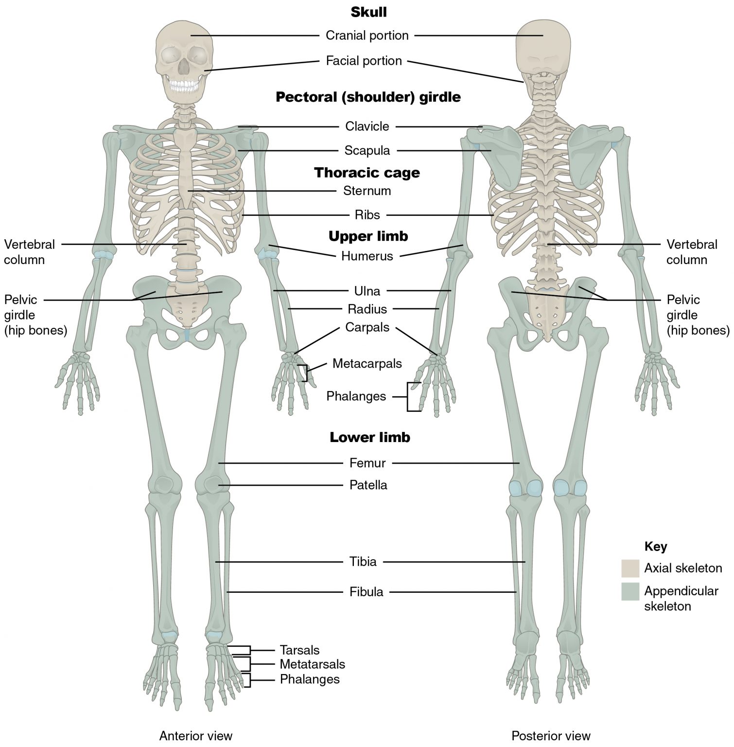 Figure \(\PageIndex{1}\): The axial skeleton consists of the skull, vertebral column, and the thoracic cage. The appendicular skeleton is made up of all bones of the upper and lower limbs.
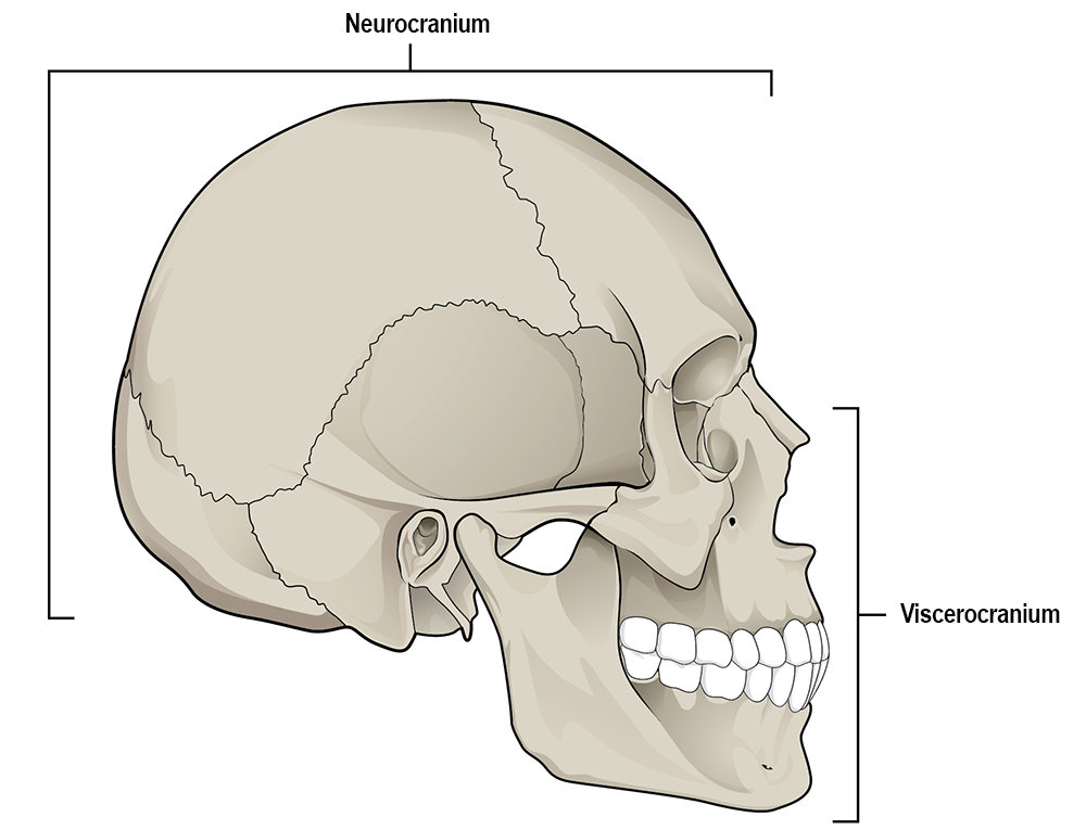 Figure \(\PageIndex{2}\): The skull consists of the cranium and the mandible. The cranium is further divided into the neurocranium and viscerocranium.
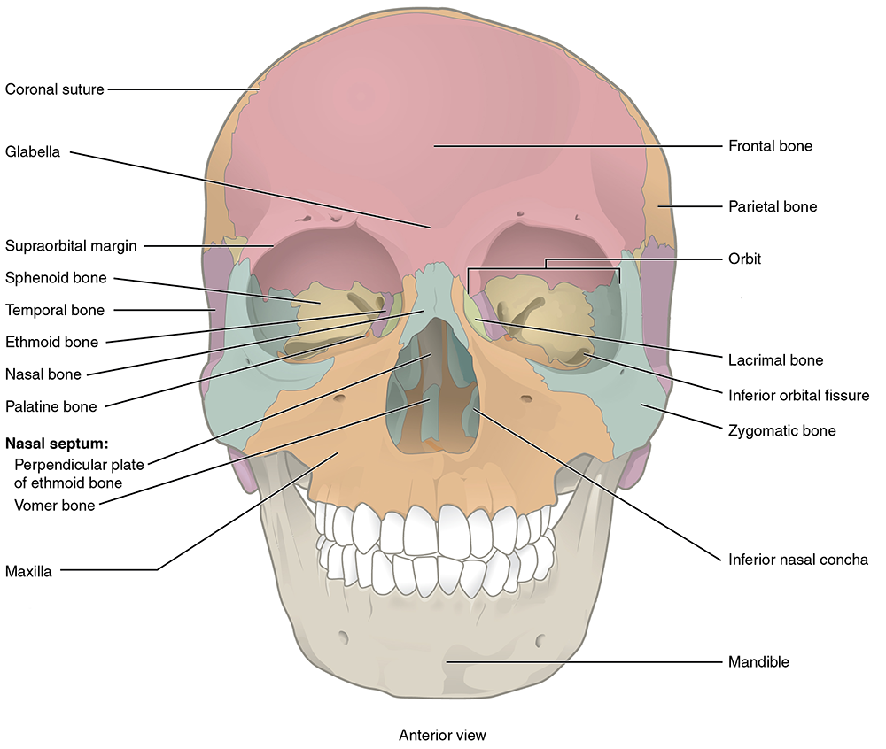 Figure \(\PageIndex{3}\): Anterior view of the skull.
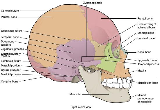 Figure \(\PageIndex{4}\): Lateral view of the skull.
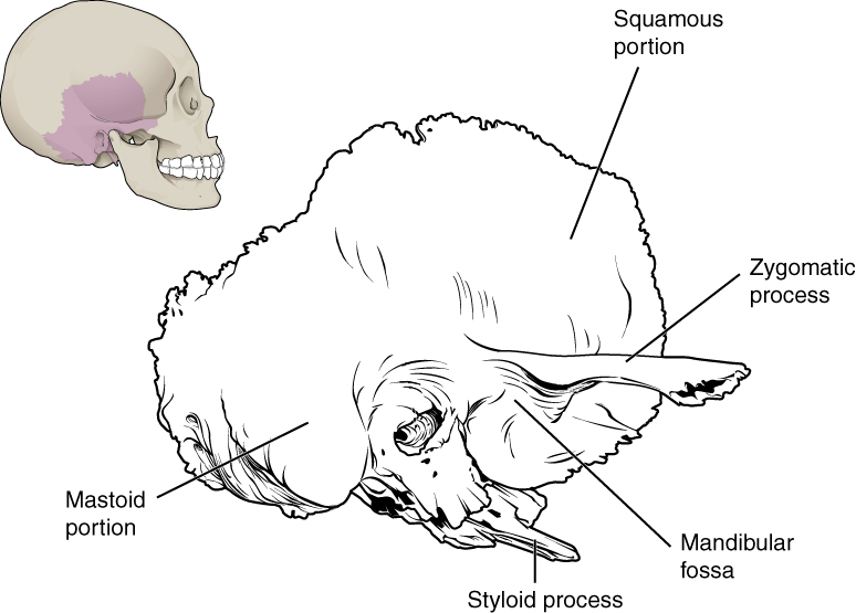 Figure \(\PageIndex{5}\): A lateral view of the isolated temporal bone shows the squamous portion.
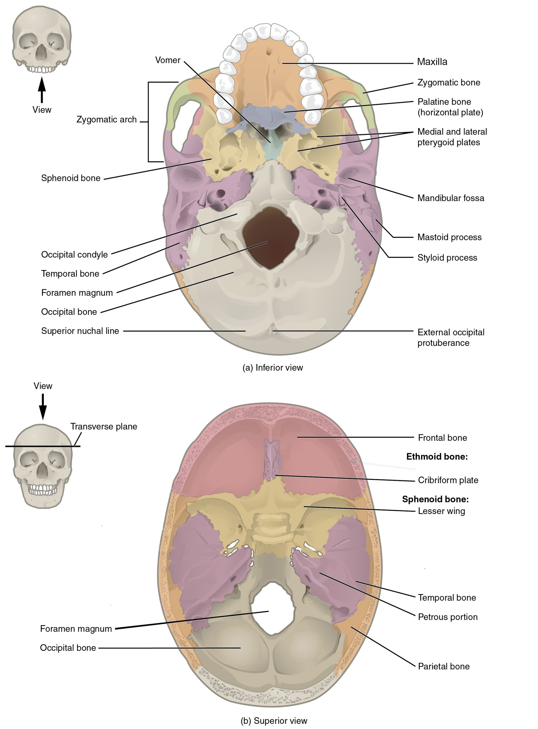 Figure \(\PageIndex{6}\): (a) The base of the cranium (b) The floor of the cranial cavity.
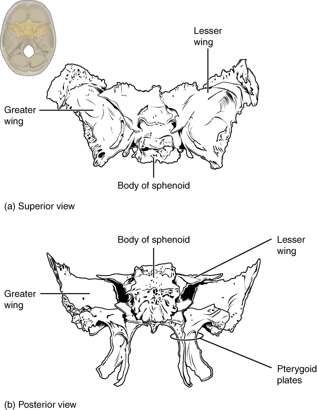 Figure \(\PageIndex{7}\): Shown in isolation in (a) superior and (b) posterior views, the sphenoid forms the central portion of the neurocranium. The sphenoid has multiple openings for the passage of nerves and blood vessels.
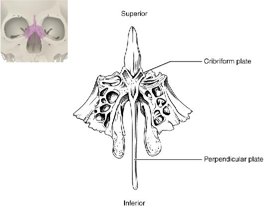 Figure \(\PageIndex{8}\): The unpaired ethmoid bone is located at the midline within the central skull. It forms the upper nasal septum and contains foramina to convey olfactory nerves.
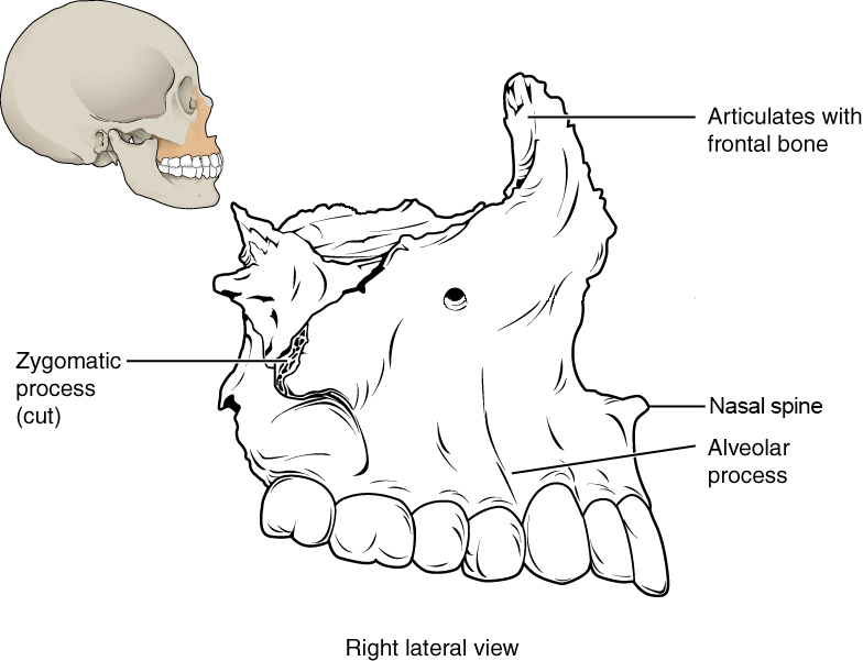 Figure \(\PageIndex{9}\): The maxilla forms the upper jaw and supports the upper teeth.
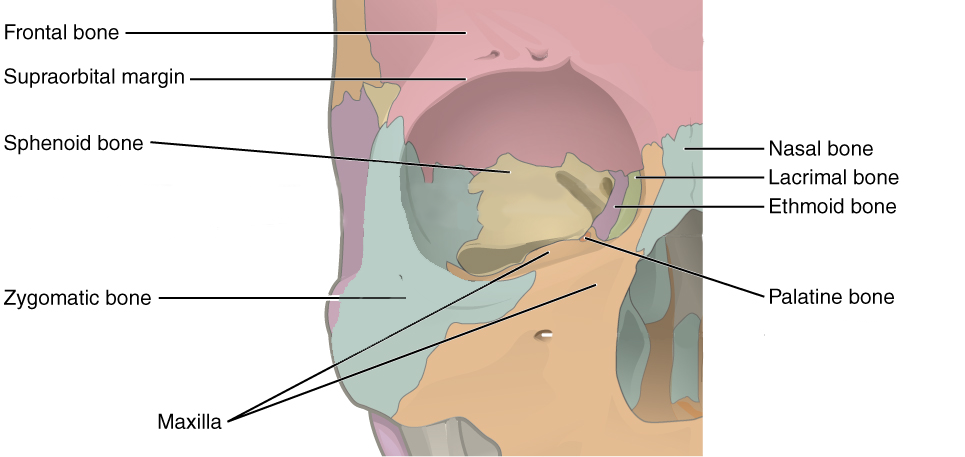 Figure \(\PageIndex{10}\): Seven skull bones contribute to the walls of the orbit: frontal, zygomatic, maxilla, lacrimal, ethmoid, palatine, and sphenoid.
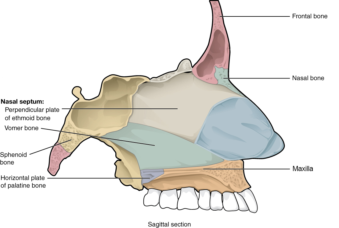 Figure \(\PageIndex{11}\): The nasal septum is formed by the perpendicular plate of the ethmoid bone and the vomer bone.
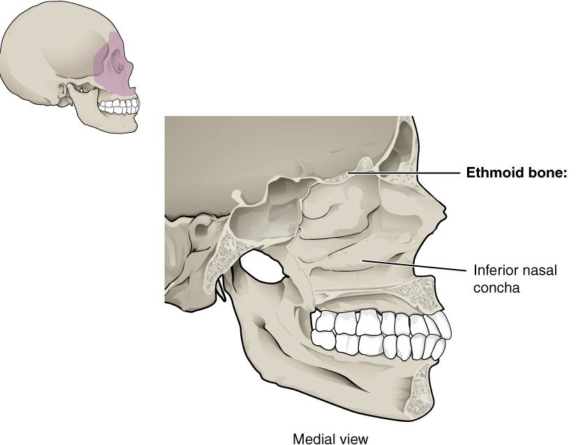 Figure \(\PageIndex{12}\): Inferior nasal concha scroll from the lateral wall of the nasal cavity.
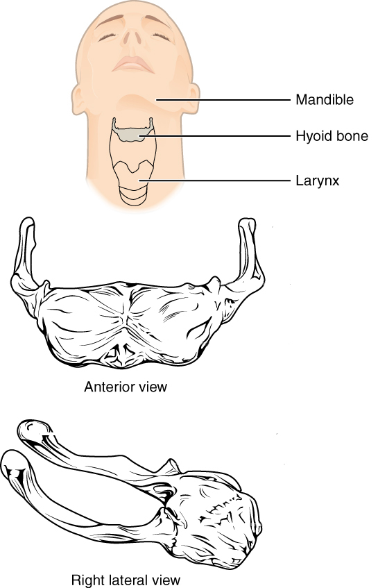 Figure \(\PageIndex{13}\): The hyoid bone is located in the upper neck and does not join with any other bone. It provides attachments for muscles that move the tongue, larynx, and pharynx.
 Figure \(\PageIndex{14}\): The mandible articulates with the temporal fossa to form the temporomandibular joint.
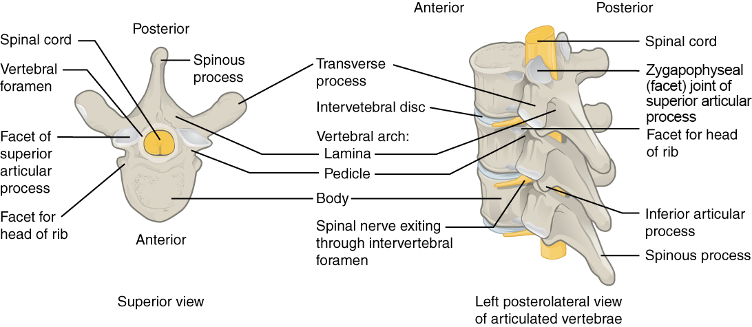 Figure \(\PageIndex{15}\): A typical vertebra consists of a body and a vertebral arch. The arch is formed by the paired pedicles and paired laminae. Arising from the vertebral arch are the transverse, spinous, superior articular, and inferior articular processes. The vertebral foramen provides for passage of the spinal cord. Each spinal nerve exits through an intervertebral foramen, located between adjacent vertebrae.
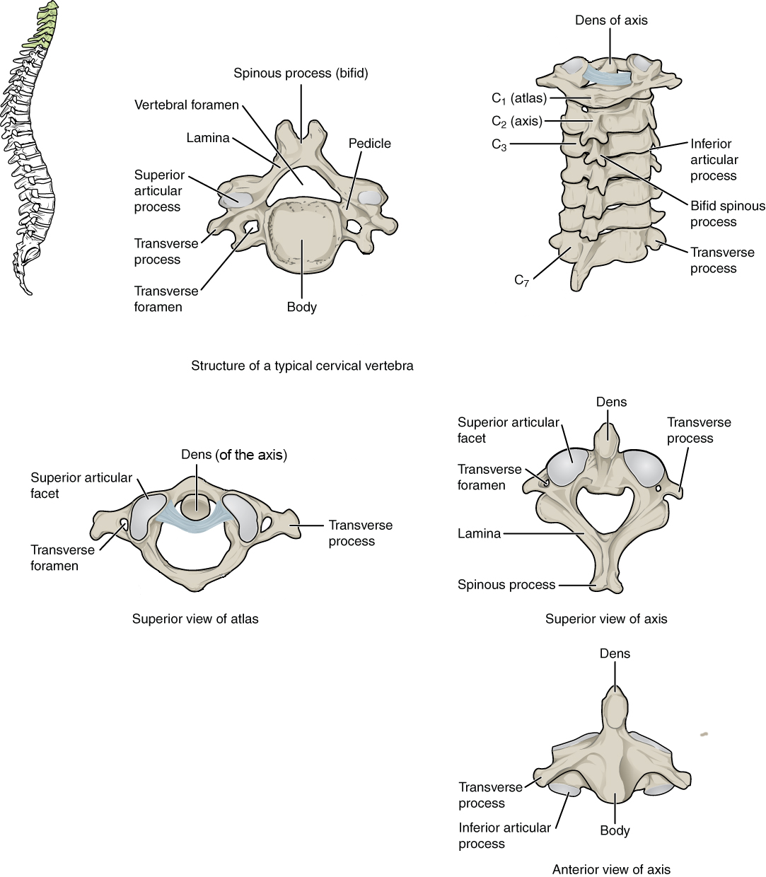 Figure \(\PageIndex{16}\): A typical cervical vertebra has a small body, a bifid spinous process, transverse processes that have a transverse foramen, and a triangular vertebral foramen. The atlas (C1 vertebra) does not have a body or spinous process. The axis (C2 vertebra) has the upward projecting dens, which articulates with the atlas.
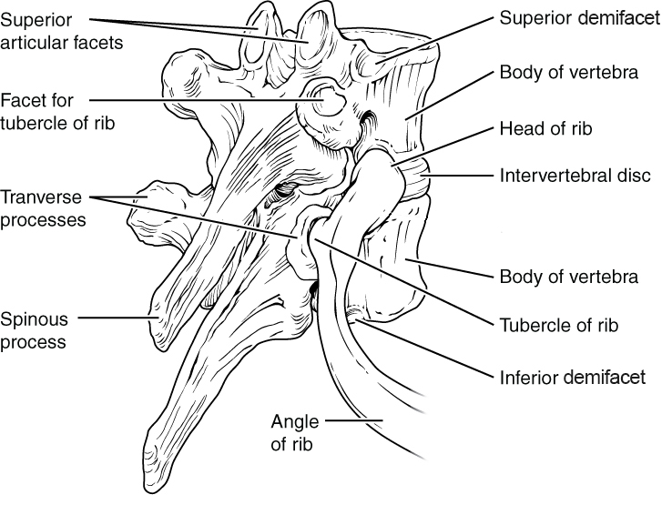 Figure \(\PageIndex{17}\): Thoracic vertebrae have superior and inferior articular facets on the vertebral body for articulation with the head of a rib, and a transverse process facet for articulation with the rib tubercle.
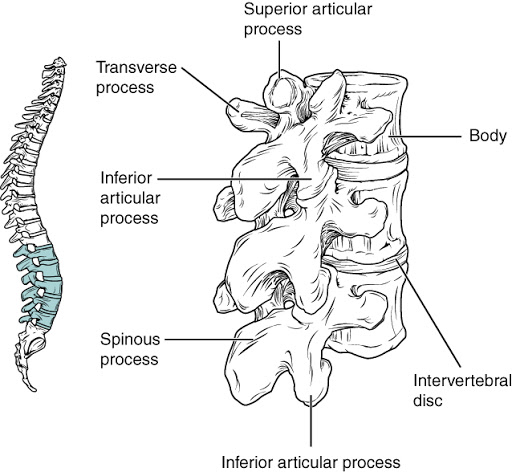 Figure \(\PageIndex{18}\): Lumbar vertebrae are characterized by having a large, thick body and a short, rounded spinous process.
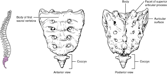 Figure \(\PageIndex{19}\): The sacrum is formed from the fusion of five sacral vertebrae, whose lines of fusion are indicated by the transverse ridges. The coccyx is formed by the fusion of three to four coccygeal vertebrae.
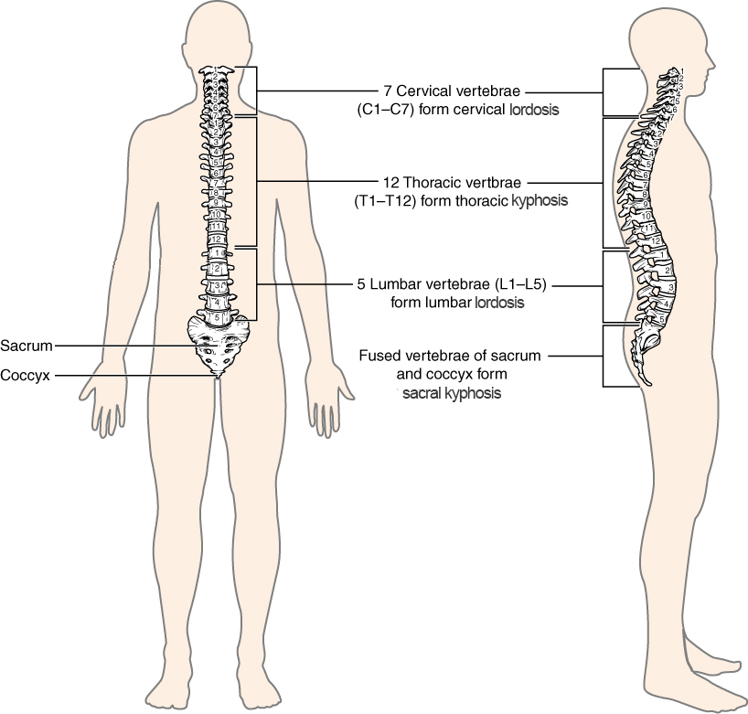 Figure \(\PageIndex{20}\): The adult vertebral column is curved in the midsagittal plane, with two primary curvatures (thoracic and sacral kyphoses) and two secondary curvatures (cervical and lumbar lordoses).
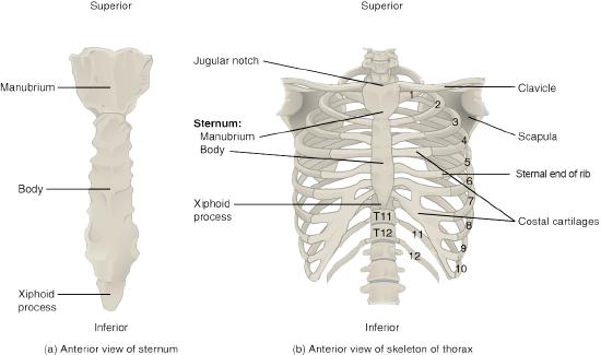 Figure \(\PageIndex{21}\): The thoracic cage is formed by the (a) sternum and (b) 12 pairs of ribs with their costal cartilages. The ribs are anchored posteriorly to the 12 thoracic vertebrae. The sternum consists of the manubrium, body, and xiphoid process. The ribs are classified as true ribs (1–7) and false ribs (8–12). The last two pairs of false ribs are also known as floating ribs (11–12).
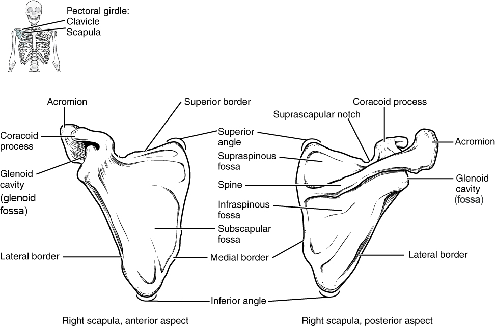 Figure \(\PageIndex{22}\): The scapula is shown from its anterior (deep) side and its posterior (superficial) side.
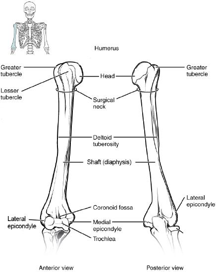 Figure \(\PageIndex{23}\): The humerus is the single bone of the upper arm region. It articulates with the radius and ulna bones of the forearm to form the elbow joint.
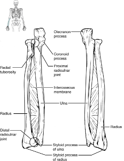 Figure \(\PageIndex{24}\): The ulna is located on the medial side of the forearm, and the radius is on the lateral side. These bones are attached to each other by an interosseous membrane.
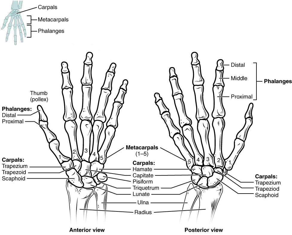 Figure \(\PageIndex{25}\): The eight carpal bones form the base of the hand. These are arranged into proximal and distal rows of four bones each. The five metacarpal bones form the palm of the hand. The thumb and fingers contain a total of 14 phalanges.
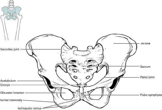 Figure \(\PageIndex{26}\): The pelvic girdle consists of two os coxae and the sacrum. It serves to anchor the axial skeleton to the lower limb.
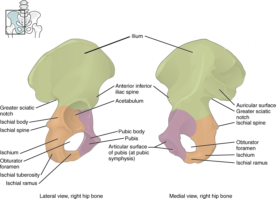 Figure \(\PageIndex{27}\): The os coxae consist of three bones that fuse during development. The ilium forms the large, fan-shaped superior portion, the ischium forms the posteroinferior portion, and the pubis forms the anteromedial portion.
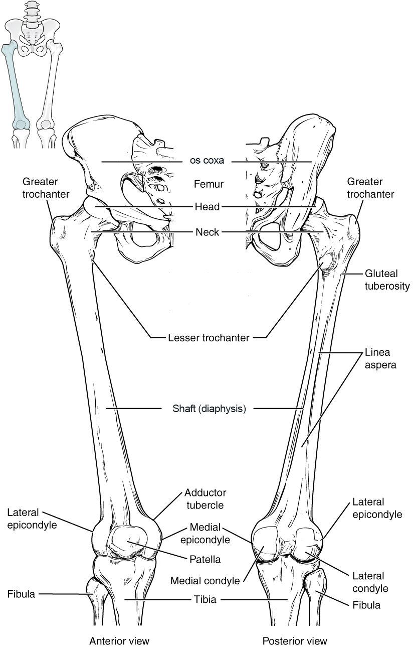 Figure \(\PageIndex{28}\): The femur is the bone of the thigh that articulates superiorly with the os coxa at the hip joint, and inferiorly with the tibia at the knee joint. The patella only articulates with the distal end of the femur.
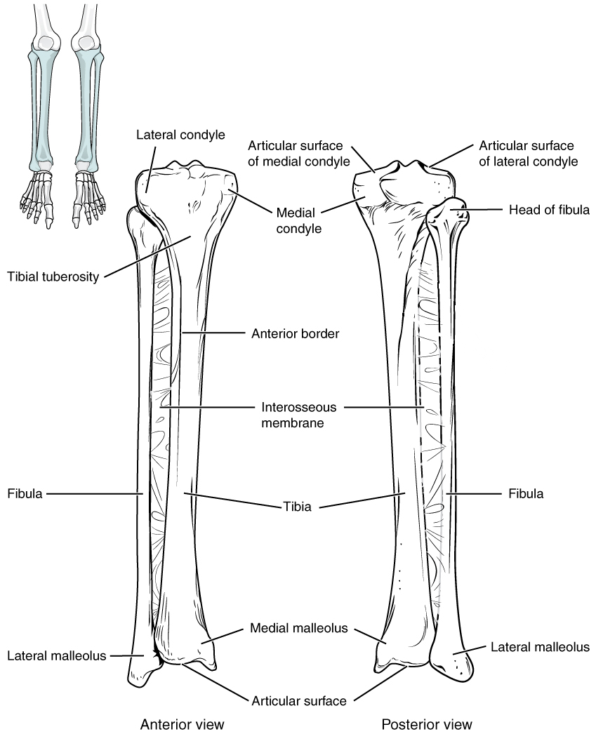 Figure \(\PageIndex{29}\): The tibia is the larger, weight-bearing bone located on the medial side of the leg. It is connected to the laterally-positioned fibula by an interosseous membrane.
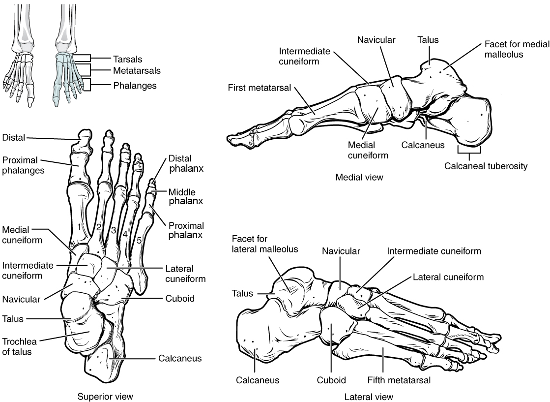 Figure \(\PageIndex{30}\): The bones of the foot are divided into three groups. The posterior foot is formed by the seven tarsal bones. The mid-foot has the five metatarsal bones. The toes contain 14 phalanges.