TERMS
- Proprioception The sense of the position of parts of the body relative to neighbouring parts of the body.
- Ventral On the front side of the human body, or the corresponding surface of an animal, usually the lower surface.
- Dorsal With respect to, or concerning the side in which the backbone is located, or the analogous side of an invertebrate.
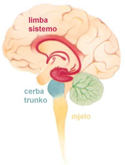
The brain’s lower-level structures consist of the brain stem and spinal cord, along with the cerebellum. With the exception of the spinal cord, these structures are largely located within the hindbrain, diencephalon (or interbrain), and midbrain. These lower dorsal structures are the oldest parts of the brain, having existed for much of its evolutionary history. As such they are geared more toward basic bodily processes necessary to survival. It is the more recent layers of the brain (the forebrain) which are responsible for the higher-level cognitive functioning (language, reasoning) not strictly necessary to keep a body alive.
The Hindbrain
The hindbrain, which includes the medulla oblongata, the pons, and the cerebellum, is responsible some of the oldest and most primitive body functions. Each of these structures is described below.
Medulla Oblongata
The medulla oblongata sits at the transition zone between the brain and the spinal cord. It is the first region that formally belongs to the brain (rather than the spinal cord). It is the control center for respiratory, cardiovascular , and digestive functions.
Pons
The pons connects the medulla oblongata with the midbrain region, and also
relays signals from the forebrain to the cerebellum. It houses the control centers for respiration and inhibitory functions. The cerebellum is attached to the dorsal side of the pons.
Cerebellum
The cerebellum is a separate region of the brain located behind the medulla oblongata and pons. It is attached to the rest of the brain by three stalks (called pedunculi ), and coordinates skeletal muscles to produce smooth, graceful motions. The cerebellum receives information from our eyes, ears, muscles, and joints about the body’s current positioning (referred to as proprioception). It also receives output from the cerebral cortex about where these body parts should be. After processing this information, the cerebellum sends motor impulses from the brain stem to the skeletal muscles so that they can move. The main function of the cerebellum is this muscle coordination. However, it is also responsible for balance and posture , and it assists us when we are learning a new motor skill, such as playing a sport or musical instrument.
Recent research shows that apart from motor functions the cerebellum also has some role in emotional sensitivity.
Human and shark brains
The shark brain diverged on the evolutionary tree from the human brain, but both still have the “old” structures of the hindbrain and midbrain dedicated to autonomic bodily processes.
The Midbrain
The midbrain is located between the hindbrain and forebrain, but it is actually part of the brain stem. It displays the same basic functional composition found in the spinal cord and the hindbrain. Ventral areas control motor function and convey motor information from the cerebral cortex. Dorsal regions of the midbrain are involved in sensory information circuits. The substantia nigra, a part of the brain that plays a role in reward, addiction, and movement (due to its high levels of dopaminergic neurons) is located in the midbrain. In Parkinson’s disease, which is characterized by a deficit of dopamine , death of the substantia nigra is evident.
The Diencephalon (“Interbrain”)
The diencephalon is the region of the embryonic vertebrate neural tube that gives rise to posterior forebrain structures. In adults, the diencephalon appears at the upper end of the brain stem, situated between the cerebrum and the brain stem. It is home to the limbic system, which is considered the seat of emotion in the human brain. The diencephalon is made up of four distinct components: the thalamus, the subthalamus, the hypothalamus, and the epithalamus.
Thalamus
The thalamus is part of the limbic system. It consists of two lobes of grey matter along the bottom of the cerebral cortex. Because nearly all sensory information passes through the thalamus it is considered the sensory “way station” of the brain, passing information on to the cerebral cortex (which is in the forebrain). Lesions of, or stimulation to, the thalamus are associated with changes in emotional reactivity. However, the importance of this structure on the regulation of emotional behavior is not due to the activity of the thalamus itself, but to the connections between the thalamus and other limbic-system structures.
Hypothalamus
The hypothalamus is a small part of the brain located just below the thalamus. Lesions of the hypothalamus interfere with motivated behaviors like sexuality, combativeness, and hunger. The hypothalamus also plays a role in emotion: parts of the hypothalamus seem to be involved in pleasure and rage, while the central part is linked to aversion, displeasure, and a tendency towards uncontrollable and loud laughing. When external stimuli are presented (for example, a dangerous stimuli), the hypothalamus sends signals to other limbic areas to trigger feeling states in response to the stimuli (in this case, fear).
The Spinal Cord
The spinal cord is a tail-like structure embedded in the vertebral canal of the spine. The adult spinal cord is about 40 cm long and weighs approximately 30 g . The spinal cord is attached to the underside of the medulla oblongata, and is organized to serve four distinct tasks:
- to convey (mainly sensory) information to the brain;
- to carry information generated in the brain to peripheral targets like skeletal muscles;
- to control nearby organs via the autonomic nervous system;
- to enable sensorimotor functions to control posture and other fundamental movements.