Identify and describe the role of the parts of the limbic system, the midbrain, and hindbrain
Areas of the Forebrain
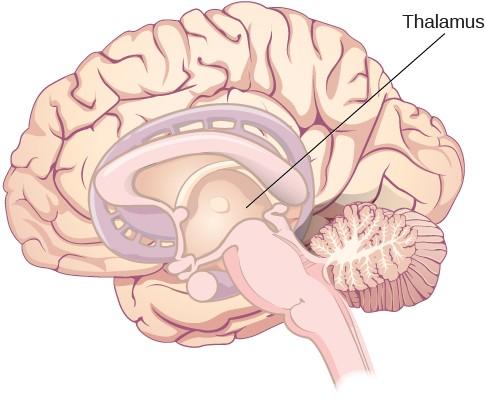
Other areas of the forebrain (which includes the lobes that you learned about previously), are the parts located beneath the cerebral cortex, including the thalamus and the limbic system. The thalamus is a sensory relay for the brain. All of our senses, with the exception of smell, are routed through the thalamus before being directed to other areas of the brain for processing (Figure 1).
Figure 9. The thalamus serves as the relay center of the brain where most senses are routed for processing.
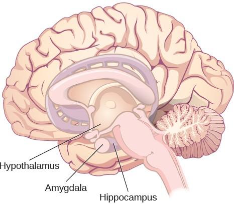
The limbic system is involved in processing both emotion and memory. Interestingly, the sense of smell projects directly to the limbic system; therefore, not surprisingly, smell can evoke emotional responses in ways that other sensory modalities cannot. The limbic system is made up of a number of different structures, but three of the most important are the hippocampus, the amygdala, and the hypothalamus (Figure 2). The hippocampus is an essential structure for learning and memory. The amygdala is involved in our experience of emotion and in tying emotional meaning to our memories. The hypothalamus regulates a number of homeostatic processes, including the regulation of body temperature, appetite, and blood pressure. The hypothalamus also serves as an interface between the nervous system and the endocrine system and in the regulation of sexual motivation and behavior.
Figure 10. The limbic system is involved in mediating emotional response and memory .
Link to Learning
Clive Wearing, an accomplished musician, lost the ability to form new memories when his hippocampus was damaged through illness. Check out the first few minutes of this documentary videofor an introduction to this man and his condition.
Midbrain and Hindbrain Structures
The midbrain is comprised of structures located deep within the brain, between the forebrain and the hindbrain. The reticular formation is centered in the midbrain, but it actually extends up into the forebrain and down into the hindbrain. The reticular formation is important in regulating the sleep/wake cycle, arousal, alertness, and motor activity.
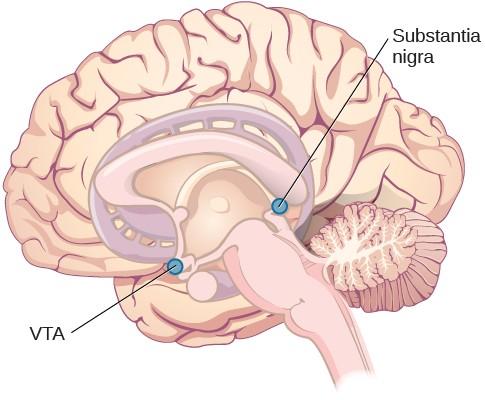
Figure 11. The substantia nigra and ventral tegmental area (VTA) are located in the midbrain.
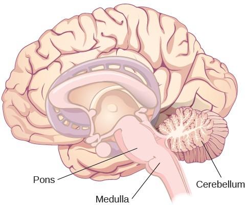
The hindbrain is located at the back of the head and looks like an extension of the spinal cord. It contains the medulla, pons, and cerebellum (Figure 4). The medulla controls the automatic processes of the autonomic nervous system, such as breathing, blood pressure, and heart rate. The word pons literally means “bridge,” and as the name suggests, the pons serves to connect the brain and spinal cord. It also is involved in regulating brain activity during sleep. The medulla, pons, and midbrain together are known as the brainstem.
Figure 12. The pons, medulla, and cerebellum make up the hindbrain.
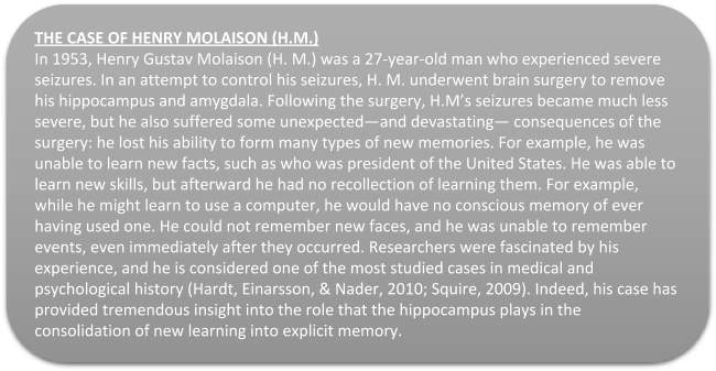
The cerebellum (Latin for “little brain”) receives messages from muscles, tendons, joints, and structures in our ear to control balance, coordination, movement, and motor skills. The cerebellum is also thought to be an important area for processing some types of memories. In particular, procedural memory, or memory involved in learning and remembering how to perform tasks, is thought to be associated with the cerebellum. Recall that H. M. was unable to form new explicit memories, but he could learn new tasks. This is likely due to the fact that H. M.’s cerebellum remained intact.
Link to Learning
Click on the link below to review each part of the brain and its purpose through the PsychSim Tutorial. The tutorial is only intended for practice. Please disregard the final screen that requests you submit answers to your instructor.
Brain and Behavior
For a fun recap of the parts of the brain, watch the following short clip from the old cartoon, Pinky and the Brain:
WHAT DO YOU THINK?: BRAIN DEAD AND ON LIFE SUPPORT
What would you do if your spouse or loved one was declared brain dead but his or her body was being kept alive by medical equipment? Whose decision should it be to remove a feeding tube? Should medical care costs be a factor?
On February 25, 1990, a Florida woman named Terri Schiavo went into cardiac arrest, apparently triggered by a bulimic episode. She was eventually revived, but her brain had been deprived of oxygen for a long time. Brain scans indicated that there was no activity in her
cerebral cortex, and she suffered from severe and permanent cerebral atrophy. Basically, Schiavo was in a vegetative state. Medical professionals determined that she would never again be able to move, talk, or respond in any way. To remain alive, she required a feeding tube, and there was no chance that her situation would ever improve.
On occasion, Schiavo’s eyes would move, and sometimes she would groan. Despite the doctors’ insistence to the contrary, her parents believed that these were signs that she was trying to communicate with them.
After 12 years, Schiavo’s husband argued that his wife would not have wanted to be kept alive with no feelings, sensations, or brain activity. Her parents, however, were very much against removing her feeding tube. Eventually, the case made its way to the courts, both in the state of Florida and at the federal level. By 2005, the courts found in favor of Schiavo’s husband, and the feeding tube was removed on March 18, 2005. Schiavo died 13 days later.
Why did Schiavo’s eyes sometimes move, and why did she groan? Although the parts of her brain that control thought, voluntary movement, and feeling were completely damaged, her brainstem was still intact. Her medulla and pons maintained her breathing and caused involuntary movements of her eyes and the occasional groans. Over the 15-year period that she was on a feeding tube, Schiavo’s medical costs may have topped $7 million (Arnst, 2003).
These questions were brought to popular conscience 25 years ago in the case of Terri Schiavo, and they persist today. In 2013, a 13-year-old girl who suffered complications after tonsil surgery was declared brain dead. There was a battle between her family, who wanted her to remain on life support, and the hospital’s policies regarding persons declared brain dead. In another complicated 2013–14 case in Texas, a pregnant EMT professional declared brain dead was kept alive for weeks, despite her spouse’s directives, which were based on her wishes should this situation arise. In this case, state laws designed to protect an unborn fetus came into consideration until doctors determined the fetus unviable.
Decisions surrounding the medical response to patients declared brain dead are complex. What do you think about these issues?
THINK IT OVER
You read about H. M.’s memory deficits following the bilateral removal of his hippocampus and amygdala. Have you encountered a character in a book, television program, or movie that suffered memory deficits? How was that character similar to and different from H. M.?
GLOSSARY
Amygdala: structure in the limbic system involved in our experience of emotion and tying emotional meaning to our memories
Cerebellum: hindbrain structure that controls our balance, coordination, movement, and motor skills, and it is thought to be important in processing some types of memory
Cerebral cortex: surface of the brain that is associated with our highest mental capabilities
Forebrain: largest part of the brain, containing the cerebral cortex, the thalamus, and the limbic system, among other structures
Hindbrain: division of the brain containing the medulla, pons, and cerebellum
Hippocampus: structure in the temporal lobe associated with learning and memory
Hypothalamus: forebrain structure that regulates sexual motivation and behavior and a number of homeostatic processes; serves as an interface between the nervous system and the endocrine system
Limbic system: collection of structures involved in processing emotion and memory
Medulla: hindbrain structure that controls automated processes like breathing, blood pressure, and heart rate
Midbrain: division of the brain located between the forebrain and the hindbrain; contains the reticular formation
Pons: hindbrain structure that connects the brain and spinal cord; involved in regulating brain activity during sleep
Reticular formation: midbrain structure important in regulating the sleep/wake cycle, arousal, alertness, and motor activity
Thalamus: sensory relay for the brain
Ventral tegmental area (VTA): midbrain structure where dopamine is produced: associated with mood, reward, and addiction
Somatosensory and Motor Cortex
Cortical Processing
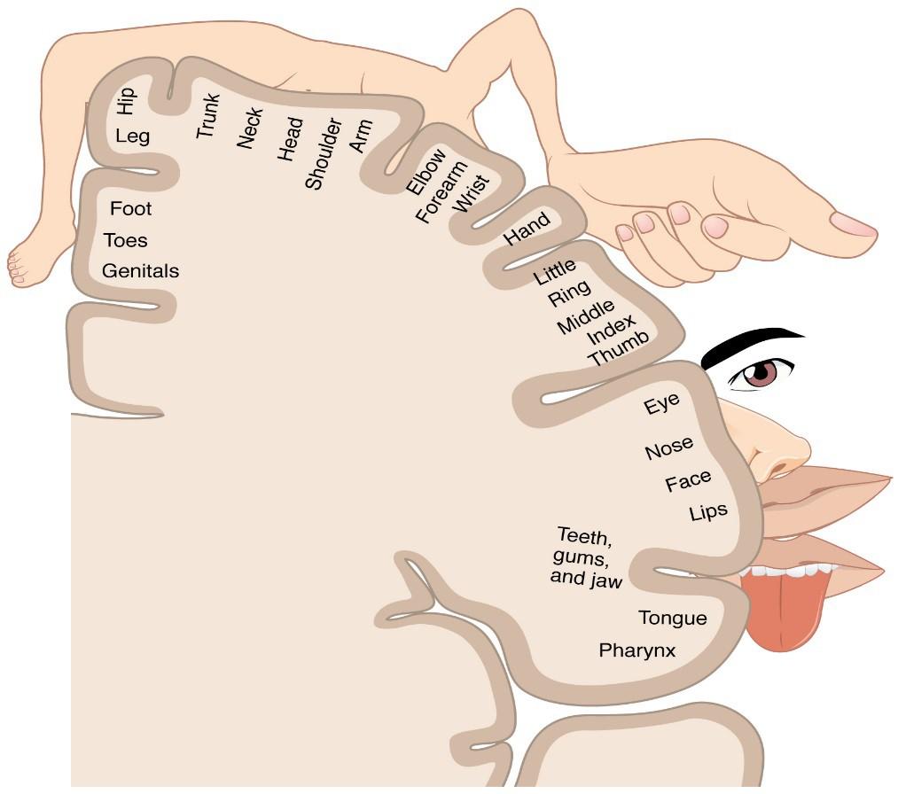
As described earlier, many of the sensory axons are positioned in the same way as their corresponding receptor cells in the body. This allows identification of the position of a stimulus on the basis of which receptor cells are sending information. The cerebral cortex also maintains this sensory topography in the particular areas of the cortex that correspond to the position of the receptor cells. The somatosensory cortex provides an example in which, in essence, the locations of the somatosensory receptors in the body are mapped onto the somatosensory cortex. This mapping is often depicted using a sensory homunculus (Figure 13).
The term homunculus comes from the Latin word for “little man” and refers to a map of the human body that is laid across a portion of the cerebral cortex. In the somatosensory cortex, the external genitals, feet, and lower legs are represented on the medial face of the gyrus within the longitudinal fissure. As the gyrus curves out of the fissure and along the surface of the parietal lobe, the body map continues through the thighs, hips, trunk, shoulders, arms, and hands. The head and face are just lateral to the fingers as the gyrus approaches the lateral sulcus. The representation of the body in this topographical map is medial to lateral from the lower to upper body. It is a continuation of the topographical arrangement seen in the dorsal column system, where axons from the lower body are carried in the fasciculus gracilis, whereas axons from the upper body are carried in the fasciculus cuneatus. As the dorsal column system continues into the medial lemniscus, these relationships are maintained. Also, the head and neck axons running from the trigeminal nuclei to the thalamus run adjacent to the upper body
fibers. The connections through the thalamus maintain topography such that the anatomic information is preserved. Note that this correspondence does not result in a perfectly miniature scale version of the body, but rather exaggerates the more sensitive areas of the body, such as the fingers and lower face. Less sensitive areas of the body, such as the shoulders and back, are mapped to smaller areas on the cortex.
Figure 13. The Sensory Homunculus. A cartoon representation of the sensory homunculus arranged adjacent to the cortical region in which the processing takes place.
The cortex has been described as having specific regions that are responsible for processing specific information; there is the visual cortex, somatosensory cortex, gustatory cortex, etc. However, our experience of these senses is not divided. Instead, we experience what can be referred to as a seamless percept. Our perceptions of the various sensory modalities—though distinct in their content—are integrated by the brain so that we experience the world as a continuous whole.
In the cerebral cortex, sensory processing begins at the primary sensory cortex, then proceeds to an association area, and finally, into a multimodal integration area. For example, somatosensory information inputs directly into the primary somatosensory cortex in the post- central gyrus of the parietal lobe where general awareness of sensation (location and type of sensation) begins. In the somatosensory association cortex details are integrated into a whole. In the highest level of association cortex details are integrated from entirely different modalities to form complete representations as we experience them.
Motor Responses
The defining characteristic of the somatic nervous system is that it controls skeletal muscles. Somatic senses inform the nervous system about the external environment, but the response to that is through voluntary muscle movement. The term “voluntary” suggests that there is a conscious decision to make a movement. However, some aspects of the somatic system use voluntary muscles without conscious control. One example is the ability of our breathing to switch to unconscious control while we are focused on another task. However, the muscles that are responsible for the basic process of breathing are also utilized for speech, which is entirely voluntary.