
8.17: Exercise- A Two-Stage Strategy for Eliminating Small But Consistent Eye Movements
- Last updated
- Save as PDF
- Page ID
- 137789

If eye movements were either large or absent, rejection of trials with eye movements would be easy. However, people may make small but consistent eye movements toward the target. Small eye movements produce small HEOG deflections, and in practice it is difficult to detect eye movements smaller than approximately 1° (16 µV) on single trials in most participants. In this exercise, we’ll see how to use a two-stage procedure (originally described by Woodman & Luck, 2003) to ensure that the data are not contaminated by small but consistent eye movements toward the target. In Stage 1, we throw out single trials with eye movements of greater than ~1°. In Stage 2, we look at the averaged HEOG-bipolar waveforms for left- and right-target trials to assess the effects of any small eye movements that remain after Stage 1.
Let’s start with Stage 1. If we’re not thoughtful about the artifact rejection parameters used at this stage, we’ll end up rejecting so many trials that we’ll need to exclude the participant. One way to minimize the number of rejected trials is to look for eye movements only until the end of the N2pc measurement window. The N2pc measurement window in the ERP CORE N2pc experiment was 200-275 ms, but to keep things simple we’ll assume a measurement window of 200-300 ms here. If the eyes move toward the target after 300 ms, this won’t impact our N2pc amplitude measurements, so we won’t reject trials with those late eye movements. This will give us more trials in our averages.
To get started on Stage 1, make sure that 15_N2pc_ICA_preprocessed_epoched is loaded and active. Select EEGLAB > ERPLAB > Artifact detection in epoched data > Step-like artifacts, and set the parameters as shown in Screenshot 8.8. We’re specifying a moving window of 200 ms, and the step function gives us the absolute value of the difference in mean voltage between the first and second halves of this window (the first 100 ms and the last 100 ms). We’re specifying a Window Step of 10, which means that we’re shifting this window in 10-ms increments. We’re specifying a test period of -200 400 rather than the entire epoch so that we don’t have to throw out trials with eye movements after the N2pc measurement window (200–300 ms).
Timing Details
The last moving window being tested will be from 200–400 ms, so an eye movement that starts at 300 ms will be detected. If we ended the test period at 300 ms, the last moving window would be from 100–300 ms, which corresponds to the difference in mean voltage between the 100–200 and 200–300 ms periods. An eye movement that began at, for example, 280 ms would only influence the voltage during the very last part of this period and would probably be missed. But this eye movement would be caught by a moving window from 180–380 ms. In general, if you are using a 200 ms moving window (which works well for eye movements), the window should end 100 ms after the time period of interest.
Screenshot 8.8
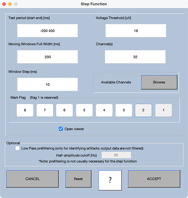
We’ve specified a threshold of 16 µV, which means that we will detect eye movements that are ~1° or larger. I like this threshold, because 1° is a nice round number, and this threshold works reasonably well with most adult participants (a higher threshold is needed for participants with noisy HEOG-bipolar signals to avoid rejecting too many trials that don’t actually have eye movements).
Go ahead and click ACCEPT to run the routine. As usual, the first thing to look at is the percentage of rejected trials. A total of 12% were rejected, which is very reasonable in terms of not reducing the signal-to-noise ratio very much. Now you should scroll through the data. (As before, I recommend displaying only the bottom 6 channels, with a vertical scale of 100 or 150.) You’ll see that the first clear eye movement, in Epoch 70, has been flagged for rejection. The blink that leaked through to the HEOG-bipolar channel in Epoch 73 was outside our window of -200 to 400 ms and was therefore not flagged, which is good. The eye movement in Epoch 78 was also outside our window and was not flagged. That’s also good, because the eye movement is too late to impact our N2pc measurement, so we want to keep this trial.
If you keep scrolling, you’ll see that the eye movements in Epochs 81, 86, and 88 were flagged. There’s a leftward (positive) eye movement in Epoch 84 that wasn’t flagged, but it was after our rejection window, so that’s good. If you go through the whole session, you’ll see that the step function did an excellent job of flagging clear eye movements that occurred during or prior to the 200–300 ms time period that we plan to use to measure N2pc amplitude.
Now let’s see what rejecting these epochs will do to the data quality. Get the table of data quality values for the data prior to flagging the artifacts and after flagging the artifacts. If you look at the aSME values for the PO7 channel in Bin 1 for the 200-300 ms time period, you’ll see that the aSME increased only slightly from the original data (0.4504) to the data excluding the marked epochs (0.4693). So, we’ve eliminated eye movements that exceeded ~1° of eye rotation without much decline in data quality. That’s good!
Now we need to implement Stage 2 of our two-stage process. Stage 2 is designed to deal with the fact that we probably failed to reject a substantial number of smaller eye movements that were directed toward the target side. These small eye movements would create a negative voltage over the contralateral hemisphere that might impact our N2pc measurements. They might also change the lateralization of the target for trials on which the target was very close to the fixation point. To assess the possibility of small but consistent eye movements remaining in the data, we need to look at the averaged HEOG-bipolar waveforms for the left-target and right-target trials.
To do this, run the averaging routine, making sure that it’s set to exclude epochs marked for rejection. Then make a left-target-minus-right-target difference wave using ERP Bin Operations. Now plot the ERPs at Channel 32 (HEOG-bipolar) for all three bins. It should look something like Screenshot 8.7C. Note that the EOG deflection is much smaller now than it was before we rejected trials with eye movements (Screenshots 8.5.A and 8.5.B), especially during the N2pc measurement window (200-300 ms).
But there is still some consistent eye movement activity in the direction of the target during this time window (i.e., the voltage is more negative on right-target trials than on left-target trials). We could try to eliminate this residual eye movement activity by decreasing our rejection threshold. However, in my experience, you can never complete remove this activity in most participants without rejecting a huge proportion of trials. For example, I tried reducing the threshold to 8 µV with the present participant, but the residual HEOG signal was quite large even though 55.4% of trials were rejected. Not surprisingly, this also increased the aSME value quite a bit. So, unless you are using a high-resolution eye tracker, you’ll always have some residual HEOG activity after artifact rejection in most participants in experiments with lateralized targets.
The question then becomes, how much residual HEOG activity can we tolerate? If we think of this question in terms of the goals described at the beginning of the chapter, we can break it into two sub-questions: 1) Is enough of the residual HEOG activity being propagated to the N2pc measurement electrodes to create a significant confound? 2) Is the amount of eye rotation implied by the residual HEOG activity large enough to create a significant change in the sensory input?
For my lab’s basic science experiments, we can afford to be extremely conservative in our answers to these questions. Our threshold for “good enough” in these experiments is a difference between left-target and right-target trials of <3.2 µV during the N2pc measurement window. In terms of eye rotation, this is an average difference in eye rotation of <0.2° between the left-target and right-target trials (which I like to think of as approximately a difference of ±0.1°). That’s a pretty tiny deviation (although we need to keep in mind that this is an average, and the deviation on single trials might be up to 1° with our 16 µV threshold). So, this seems “good enough” in terms of the change to the sensory input.
We ordinarily measure the N2pc at all of the parietal and occipital electrode sites, and the propagation factor is 3% or less from the HEOG-bipolar sites to each of these sites (according to Lins et al., 1993). Thus, a voltage difference of <3.2 µV at HEOG-bipolar corresponds to a voltage difference of <0.1 µV at the sites where we are measuring the N2pc component. That seems “good enough” in terms of any confounding voltage in our N2pc measurements.
Have we succeeded in meeting this 3.2 µV criterion in the present participant? It’s difficult to be sure in the current plot. A convenient way to see if we’ve met the criterion is to plot the difference wave using a Y range of -3.2 to +3.2 µV and a time range of -200 to +300 ms. Go ahead and do this. The result should look something like Screenshot 8.7D. If the voltage ever exceeded 3.2 µV, the waveform would be “clipped off” in the plot. Although the waveform did get near the top of this voltage range near the end of the 200-300 ms N2pc measurement window, it never exceeded this threshold. In other words, the small amount of residual eye movement activity for this participant meets our criterion for “good enough.”
What should you do if the residual eye movement activity for a given participant is >3.2 µV? The first step is to try changing the rejection parameters. Most obviously, you can try reducing the rejection threshold. Sometimes changing the rejection time window can also help. Your goal is to see if you can reduce the residual HEOG activity to <3.2 µV in the N2pc measurement window (or the measurement window for whatever component you’re studying) without rejecting too many trials.
As mentioned earlier in this chapter, my lab automatically excludes participants in our basic science experiments if more than 25% of trials are rejected (which includes trials rejected for other reasons, such as blinks). If we can’t get the residual HEOG under 3.2 µV without rejecting more than 25% of trials, we exclude the participant from the final analyses. We’ve used this approach for about 30 years, and it has worked very well. We end up excluding approximately 20% of participants, which is tolerable. In our schizophrenia studies, we find that both the patient and control groups make more eye movements than the college-age participants in our basic science studies, so we double our thresholds. That is, we require that the residual HEOG activity is <6.4 µV, and we exclude participants for whom more than 50% of trials were rejected.
What thresholds should you use in your own analyses? I can’t answer that question, because it depends on the nature of your research. As always, your choice should be made on the basis of the fundamental goal of increasing your ability to accurately answer the scientific question that your studies are designed to address. And you might use aSME values rather than the percentage of trials rejected to decide whether too many trials have been rejected for a given participant. Whatever criteria you choose, however, it is extremely important that those criteria are set prior to analyzing the data.
I hope that these N2pc exercises have provided you with a clear procedure for minimizing eye movement artifacts in experiments with lateralized targets or responses. But even more, I hope these exercises serve as a good example of how to conceptualize the goals of artifact rejection and how to achieve those goals.