
2.1: Introduction to Research in Biological Psychology
- Page ID
- 110460
This page is a draft and under active development. Please forward any questions, comments, and/or feedback to the ASCCC OERI (oeri@asccc.org).
- Differentiate between the purpose of structural and functional imaging.
- Describe the differences between spatial resolution and temporal resolution.
Overview
This chapter will describe the various ways that biological psychologists study the brain. There are many ways to categorize the techniques that are used when studying the brain. We will start by covering the non-invasive techniques, where we are able to study the brain without getting direct physical access to the brain (think of fixing a broken pipe in a wall without having to open the wall up). Then we will move into the invasive techniques, where we study the brain by having direct access (an example would be fixing a broken pipe in a wall by tearing a hole in the wall). Then we will discuss various neuropsychological techniques, where we learn about the brain using people with some sort of brain “issue.” For example, people with epilepsy have been extensively studied and we can learn a lot about how the brain works from them. Finally, the last section will address ethical considerations of biological psychology research.
In this section, we will start with a discussion of many modern day neuroscience terms that are important for understanding the different techniques in future sections.
The Terminology of Modern Research Techniques
We have come a long way since Phineas Gage with how we study the brain. Many techniques now allow us to understand how the brain works without waiting for a horrific accident to occur or conducting some sort of surgery (although, as you will see, we still use surgical techniques to study the brain). Techniques have been developed that allow us to see what the brain looks like, as a still image (structurally) or in action (functionally).
Structural Imaging
Structural imaging techniques are useful in many situations such as locating tumors, sites of physical brain damage, or finding size differences between the structures of the brain between various groups. Magnetic resonance imaging (MRI), for example, is one such technique that is commonly used to study the brain and to diagnosis knee and shoulder injuries. Structural imaging techniques allow us to look inside the brain (or body) without having to go inside.
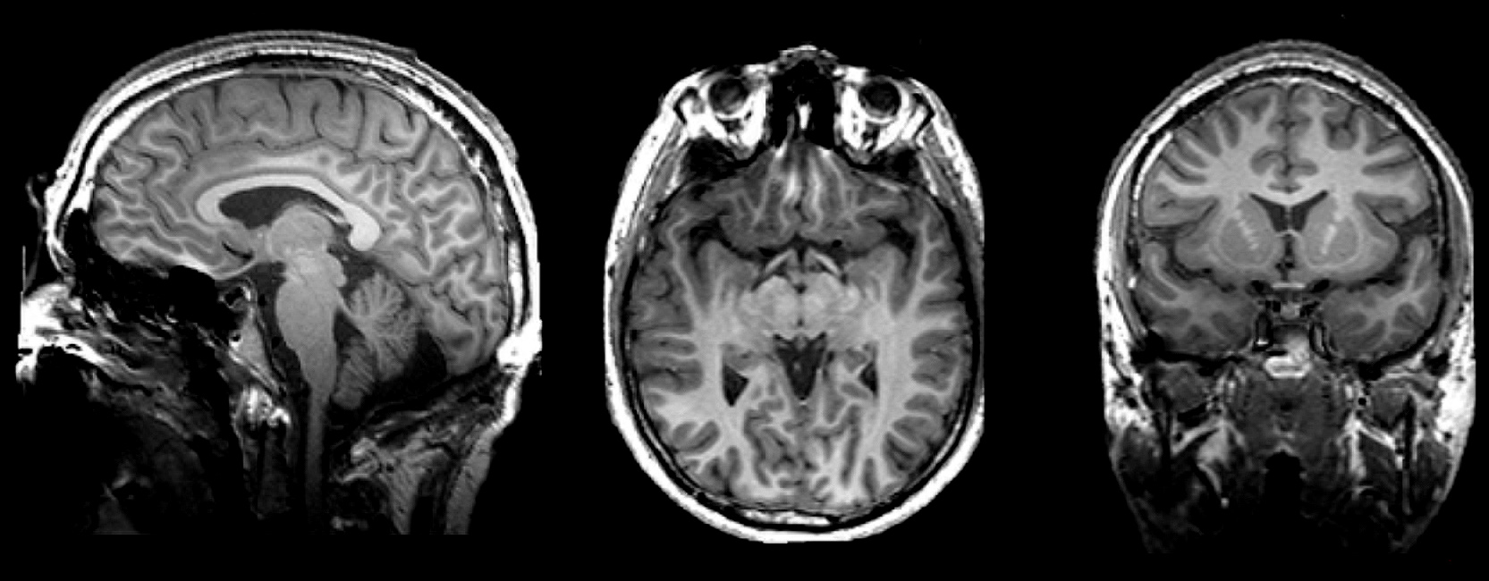
A series of MRI images can be used to create a picture of the brain.
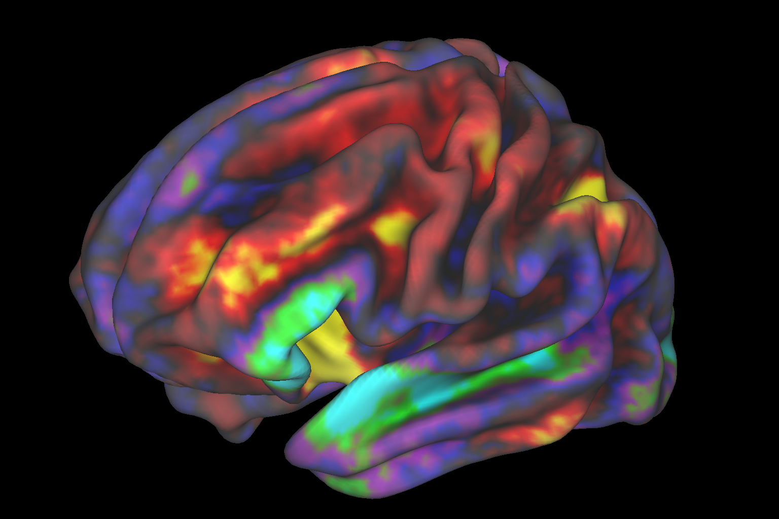
Functional Imaging
Many researchers are also interested in how the brain works. Some studies begin with the scientific question of “what does this part do?” Or more commonly, “Where in the brain does this happen?” Functional imaging techniques allow researchers to learn about the brain activity during various tasks by creating images based on the electrical activity or the absorption of various substances that occurs while a subject is engaging in a task. Such techniques can be used, for example, to visualize the parts of the brain that respond when we're exposed to stimuli that upset us or make us happy.
Temporal Versus Spatial Resolution
Within functional imaging techniques, researchers are frequently focused on one of two questions. They may ask “When does this activity occur?” Or “Where does this activity occur?” Some techniques are better for answering one of these questions, whereas other techniques are better for answering the other question. We describe how well a technique can determine when the activity has occurred as temporal resolution. For example, was the brain region activity occurring sometime in the last hour, the last minute, the last second, or within milliseconds? While some techniques are excellent at determining precisely when the activity occurred and other techniques are quite terrible at it. Additionally, we can describe how well a technique can determine where the activity has occurred as spatial resolution. For example, did the activity occur in the temporal lobe somewhere or can we narrow that down to a specific gyrus (ridge) or sulcus (groove) of the cerebral cortex? If it occurred on a particular gyrus can we narrow it down to a particular portion of that gyrus? As with temporal resolution, some techniques are excellent at determining precisely where the activity occurred whereas other techniques are less accurate.
Summary
How we study the brain has come a long way since the days of Phineas Gage. Although, as we will discuss, we still learn about the brain from accidents and other traumatic brain events, we have a variety of other techniques that we can use now to study the brain in healthy individuals. These techniques allow us to answer questions about what the brain looks like, what specific parts do, and when they do it.
Attributions
- Psychophysiological Methods in Neuroscience by Zachary Infantolino and Gregory A. Miller is licensed under a CC BY-NC-SA 4.0 International License.
- Figure \(\PageIndex{1}\): "Deadstar's brain" by Daniele Oberti is licensed under CC BY-NC-ND 2.0
- Figure \(\PageIndex{2}\): "fMRI Image of Preteen Brain" by NIH Image Gallery is licensed under CC BY-NC 2.0