
13.1: Sex Hormones, Sexual Motivation, and Reproductive Behaviors
- Page ID
- 130098
This page is a draft and under active development. Please forward any questions, comments, and/or feedback to the ASCCC OERI (oeri@asccc.org).
- Distinguish between the organizational and activational effects of sex hormones
- Describe some reciprocal interactions between sex hormones and reproductive behavior
- Understand basic biological mechanisms regulating sexual behavior and motivation
- Identify the role of the brain in responding to sexual stimuli
- Explain the role of hormones in parental behavior
Overview
This module discusses the relationship between sex and hormones (including organizational and activational effects, maturation of the reproductive systems, interactions between hormones and behavior, and anabolic steroids), sexual behavior as a form of motivation (including physiological mechanisms and human sexual behavior and motivation), sex and the brain (including erogenous zones, and the role of the hypothalamus and the pituitary gland), and parental behavior (including hormones and rodent maternal behavior, maternal aggression, and hormones and human maternal behavior).
Human Sexuality in Context
Human sexuality refers to people's sexual interest in and attraction to others, as well as their capacity to have erotic experiences and responses. Sexuality may be experienced and expressed in a variety of ways, including (but not limited to) thoughts, desires, practices, roles, and behaviors. This chapter mostly focuses on the biological and physical aspects of sexuality- human reproductive anatomy and functions, including the human sexual response cycle and the basic biological drive that exists in all species- but also addresses the psychological domains of gender identity and sexual orientation. We begin with hormones, as they play such an integral role in the development and expression of human sexuality.
Sex and Hormones
Because of their inextricable link to the development and maturation of reproductive anatomy (both prenatally and during puberty), as well as the promotion and maintenance of adult reproductive functions, Biological Psychology textbooks often include hormones in the chapter on sex and reproductive behaviors. However, many other bodily functions (such as growth, sleep, hunger, satiation, and maintenance of blood pressure, to name a few) are regulated by hormones, so we have opted to cover basics of the endocrine system (the organs which produce and secrete hormones) in the Nervous System Anatomy chapter. Please refer to that chapter if you need a brief overview of hormones and the endocrine system.
Organizational and Activational Effects of Sex Hormones
Although the dichotomy cannot be strictly applied, the effects of steroid sex hormones have typically been characterized as organizational versus activational. Organizational effects result in permanent changes that usually occur early in development, such as the prenatal development of human reproductive structures (also known as primary sex characteristics- differences in male and female bodies that are present at birth). Activational effects are usually temporary and occur throughout life, dependent on specific conditions. Thus, structures in the body, brain, and nervous system are thought to be organized (in a male-typical or female-typical manner) through the action of steroid hormones early in development, and then later on activated by steroid hormones, resulting in structures and behaviors that differ between the sexes (Arnold and Breedlove, 1985). While this simple dichotomy works well with animals with very distinct sexual dimorphism in behavior (such as rats, where only males attempt to mount other rats and only females exhibit a sexually receptive posture called lordosis), it cannot be applied in such a straightforward manner to people. For one thing, humans do not exhibit any sexual behaviors that are strictly stereotyped and unique to either males or females. Nonetheless, some of the differences between human males and females can be attributed, at least in part, to either the organizational or activational effects of sex hormones.
One example of a permanent organizational effect is the difference between males and females in the secretion of certain hormones (related to reproductive function) by the hypothalamus; adult females follow a pattern of cyclical hormone release (related to their fertility cycles), whereas males do not. Changing the hormones that are present in the body later in life will not change the capacity of the hypothalamus to cycle the release of these hormones. Some secondary sex characteristics (changes that occur in male and female bodies during the sexual maturation that occurs with puberty) are also permanent changes- requiring surgery to alter- such as the development of breasts and wider hips in women or a prominent Adam's apple (cartilage around the thyroid in the neck area) and broader shoulders in men. In contrast, some secondary sex characteristics, like the presence of facial hair in men (but not in women), can be modified with changes to hormone levels in the adult body of either sex.
One example of a temporary activational effect is lactation, or the production and secretion of milk from the mammary glands (breasts). Normally this only occurs in females after childbirth, when the necessary combination of the hormones estrogen, progesterone, and prolactin are present. "However, if a man is treated with this hormone combination, his mammary glands not only can produce milk, but he can nurse a baby! Thus, lactation is an "activated-only" action of hormones, and there is no organized sex difference in the mammary gland tissue itself." (Jones and Lopez, 2006, page 466). So, while modern science cannot yet assist transgender women (whose biological birth sex was male) in carrying and giving birth to an infant, they can nonetheless participate in nursing the baby.
Sex Hormones and Maturation
The main categories of sex hormones are androgens (more prevalent in biological males), of which testosterone is a primary example, and estrogens (more prevalent in biological females), of which estradiol is a primary example. It is important to note that all individuals have both androgens ("male hormones") and estrogens ("female hormones"), but the relative proportion of the hormones present in a given body varies, usually correlated with biological sex. Testosterone is secreted by the testes in males and in small amounts by the ovaries in females (although most is converted to estradiol). A small amount of testosterone is also secreted by the adrenal glands (endocrine glands positioned on top of the kidneys) in both sexes. Estrogens are secreted by the ovaries in females and by fat cells in both sexes.
Male and female reproductive systems are different at birth (due to differences in primary sex characteristics), but the gonads (testes in males and ovaries in females) are immature and incapable of producing gametes (sperm in males and eggs in females) or sex hormones. Maturation of the reproductive system occurs during puberty when hormones from the hypothalamus and pituitary gland stimulate the testes or ovaries to start producing sex hormones again. Sex hormones, in turn, lead to the growth and maturation of the reproductive organs, rapid body growth, and the development of secondary sex characteristics, such as pubic and underarm hair in both sexes, facial hair in males and breasts in females.
Interactions Between Sex Hormones and Reproductive Behaviors
As discussed in Chapter 4.6, the interaction between hormones and behavior is bidirectional: hormones can influence behavior, and behavior can sometimes influence hormone concentrations. Hormones travel through the blood, influencing the nervous system to regulate an individual's behaviors, some of which are related to sexuality and reproduction (such as aggression, mating, and parenting). Some hormone-behavior interactions that are related specifically to reproductive behaviors are described below.
Hormonal Influence on Reproductive Behaviors
Hormones coordinate the physiology and behavior of individuals. Over evolutionary time, hormones have been co-opted by the nervous system to influence behavior to ensure reproductive success. For example, the same hormones, testosterone and estradiol, that cause gamete (egg or sperm) maturation also promote mating behavior. This dual hormonal function ensures that mating behavior occurs when animals have mature gametes available for fertilization. Similarly, during pregnancy estrogens and progesterone concentrations are elevated, and these hormones are also involved in maternal behavior in the mothers.
How might hormones affect behavior? While hormones do not cause behavioral changes, they influence the components that interact to produce behavior- sensory systems (input), the central nervous system (integration), and muscles and glands (output). In this manner, specific stimuli are more likely to elicit certain responses in the appropriate behavioral or social context. In other words, hormones change the probability that a particular behavior will occur in the appropriate situation (Nelson, 2011). This is a critical distinction that can affect how we think of hormone-behavior relationships. In most cases, hormones can be considered to affect behavior by influencing any or all of these components.
An example of the influence of hormones on a simple behavior is singing in zebra finches. Only male zebra finches sing. If the testes of adult male finches are removed, then the birds reduce singing, but castrated finches resume singing if the testes are reimplanted, or if the birds are treated with either testosterone or estradiol. Although we generally consider androgens to be “male” hormones and estrogens to be “female” hormones, it is common for testosterone to be converted to estradiol in nerve cells. Thus, many male-like behaviors are associated with the actions of estrogens! Singing behavior is most frequent when blood testosterone or estrogen concentrations are high. Males sing to attract mates or ward off potential competitors from their territories.
Behavioral Influence on Sex Hormones
How might behaviors affect hormones? If a male mouse or rhesus monkey loses a fight, blood testosterone levels decrease for several days or even weeks afterward. Comparable results have also been reported in humans. Testosterone concentrations are affected not only in humans involved in physical combat, but also in those involved in simulated battles. For example, testosterone concentrations were elevated in winners and reduced in losers of regional chess tournaments.
People do not have to be directly involved in a contest to have their hormones affected by the outcome of the contest. Male fans of both the Brazilian and Italian teams were recruited to provide saliva samples to be assayed for testosterone before and after the final game of the World Cup soccer match in 1994. At the end of the game, Brazil and Italy were tied for regulation and overtime, but then Brazil won in penalty shots. The Brazilian fans were elated and the Italian fans were crestfallen. When the samples were assayed, 11 of 12 Brazilian fans who were sampled had increased testosterone concentrations, and 9 of 9 Italian fans had decreased testosterone concentrations, compared with pre-game baseline values (Dabbs, 2000).

In some cases, hormones can be affected by anticipation of behavior (Figure \(\PageIndex{1}\)). For example, testosterone concentrations also influence sexual motivation and behavior in women. In one study, the interaction between sexual intercourse and testosterone was compared with other activities (cuddling or exercise) in women (van Anders, Hamilton, Schmidt, & Watson, 2007). On three separate occasions, women provided a pre-activity, post-activity, and next-morning saliva sample. After analysis, the women’s testosterone was determined to be elevated prior to intercourse as compared to other times. Thus, an anticipatory relationship exists between sexual behavior and testosterone. Testosterone values were higher post-intercourse compared to exercise, suggesting that engaging in sexual behavior may also influence hormone concentrations in women.
Anabolic Steroids
The endocrine system can be exploited for illegal or unethical purposes. A prominent example of this is the use of steroid drugs by professional athletes. Commonly used for performance enhancement, anabolic steroids are synthetic versions of the sex hormone testosterone. By boosting natural levels of this hormone, athletes experience increased muscle mass. Synthetic versions of human growth hormone are also used to build muscle mass.
The use of performance-enhancing drugs is banned by all major collegiate and professional sports organizations in the United States because they impart an unfair advantage to athletes who take them. In addition, the drugs can cause significant and dangerous side effects. For example, anabolic steroid use can increase cholesterol levels, raise blood pressure, and damage the liver. Altered testosterone levels (both too low or too high) have been implicated in causing structural damage to the heart, and increasing the risk for cardiac arrhythmias, heart attacks, congestive heart failure, and sudden death. Paradoxically, steroids can cause shriveled testes and enlarged breast tissue in men. In females, their use can cause analogous effects such as an enlarged clitoris and growth of facial hair. In both sexes, their use can promote increased aggression (commonly known as “roid-rage”), depression, sleep disturbances, severe acne, and infertility.
Sexual Behavior as a Form of Motivation
Sex is an important part of most people's lives. From an evolutionary perspective, the reason is obvious—perpetuation of the species. Sexual behavior in humans, however, involves much more than reproduction. Sexual arousal is the drive state that results in thoughts and behaviors related to sexual activity. It is generated by a large range of internal and external mechanisms that are triggered either after the extended absence of sexual activity or by the immediate presence and possibility of sexual activity (or by cues commonly associated with such possibilities). This section provides an overview of some research that has been conducted on sexual behavior and motivation.
Sexual motivation, often referred to as libido, is a person's overall sexual drive or desire for sexual activity. This motivation is determined by biological, psychological, and social factors. In most mammalian species, sex hormones control the ability to engage in sexual behaviors. However, sex hormones do not directly regulate the ability to have sexual intercourse in primates (including humans); rather, they are only one influence on the motivation to engage in sexual behaviors. Social factors, such as work, family, and relationship issues also have an impact, as do internal psychological factors like personality and lifestyle stress. Sex drive may also be affected by medical conditions (including illness or injury), medications, and pregnancy.
Physiological Mechanisms of Sexual Motivation and Behavior
Much of what we know about the physiological mechanisms that underlie sexual motivation and behavior comes from animal research. Research on male rats suggests that limbic system structures such as the amygdala and nucleus accumbens are especially important for sexual motivation. Damage to these areas results in a decreased motivation to engage in sexual behavior, while leaving the ability to do so intact (Everett, 1990; Figure \(\PageIndex{2}\)). Similar dissociations of sexual motivation and sexual ability have also been observed in the female rat (Becker, Rudick, & Jenkins, 2001; Jenkins & Becker, 2001).
The hypothalamus plays an important role in motivated behaviors, and sex is no exception. In fact, lesions to an area of the hypothalamus called the medial preoptic area completely disrupt a male rat’s ability to engage in sexual behavior, but, surprisingly, do not change how hard a male rat is willing to work to gain access to a sexually receptive female (Figure \(\PageIndex{3}\)). This suggests that the ability to engage in sexual behavior and the motivation to do so may be mediated by neural systems distinct from one another.
Human Sexual Motivation and Behavior
Vasopressin is also involved in the male arousal phase, and the increase of vasopressin during erectile response may be directly associated with increased motivation to engage in sexual behavior. The relationship between hormones and female sexual motivation is not as well understood, largely due to the overemphasis on male sexuality in Western research. Estrogen and progesterone typically regulate motivation to engage in sexual behavior for females, with estrogen increasing motivation and progesterone decreasing it. The levels of these hormones rise and fall throughout a woman's menstrual cycle. Research suggests that testosterone, oxytocin, and vasopressin are also implicated in female sexual motivation in similar ways as they are in males, but more research is needed to understand these relationships.
Sex and the Brain
The brain is the organ that translates the nerve impulses from the skin into pleasurable sensations. It controls nerves and muscles used during sexual behavior. The brain regulates the release of hormones, which are believed to be the physiological origin of sexual desire. The cerebral cortex (the outer layer of the brain that allows for thinking and reasoning) is believed to be the origin of sexual thoughts and fantasies. Deep to the cortex is the limbic system, a collection of structures believed to be the origin of emotions and feelings, also important for sexual behavior. The limbic system includes the amygdala, hippocampus, cingulate gyrus, and septal nucleus. The septal nucleus, an area that receives reciprocal connections from many other brain regions (including the hypothalamus and the amygdala), seems to play an important role in sexual pleasure. This region shows rhythmic spiking activity during sexual orgasm, and is also one of the brain regions that rats will most reliably voluntarily self-stimulate (Olds & Milner, 1954). In humans, placing a small amount of acetylcholine into this region, or stimulating it electrically, has been reported to produce a feeling of imminent orgasm (Heath, 1964; Heath, 1972).
Erogenous Zones
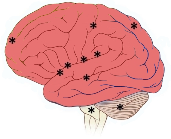
At first glance- or touch for that matter- the glans of the clitoris and penis are the parts of our anatomies that seem to bring the most pleasure. However, these two organs pale in comparison to our central nervous system’s capacity for pleasure. Extensive regions of the brain and brainstem are activated when a person experiences pleasure, including the insula, temporal cortex, limbic system, nucleus accumbens, basal ganglia, superior parietal cortex, dorsolateral prefrontal cortex, and cerebellum (Ortigue et al., 2007; Figure \(\PageIndex{4}\)). Neuroimaging techniques show that these regions of the brain are active when patients have spontaneous orgasms involving no direct stimulation of the skin (e.g., Fadul et al., 2005) and when experimental participants self-stimulate erogenous zones (e.g., Komisaruk et al., 2011). Erogenous zones are particularly sensitive areas of skin (which may be different across individuals), and (like all body areas with sensation) are connected (via the nervous system) to the somatosensory cortex in the brain (refer to the section on the central nervous system).
A study by Nummenmaa and his colleagues (2016) used a unique method to test the hypothesis that the more sensitive areas of our bodies have greater potential to evoke pleasure. The Nummenmaa research team showed experimental participants images of same- and opposite-sex bodies. They then asked the participants to color the regions of the body that, when touched, they or members of the opposite sex would experience as sexually arousing while masturbating or having sex with a partner. Nummenmaa found the expected “hotspot” erogenous zones around the external sex organs, breasts, and anus, but also reported areas of the skin beyond these hotspots: “[T]actile stimulation of practically all bodily regions trigger sexual arousal….” Moreover, he concluded, “[H]aving sex with a partner…”—beyond the hotspots—“…reflects the role of touching in the maintenance of…pair bonds.” This also underlines the fact that individuals are different, and erogenous zones vary correspondingly.
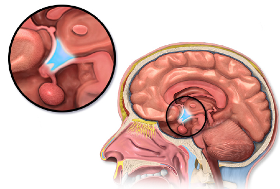
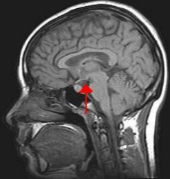
The Role of the Hypothalamus and the Pituitary Gland
Parental Behavior
Parental behavior can be considered to be any behavior that contributes directly to the survival of fertilized eggs or offspring that have left the body of the female. There are many patterns of parental care in mammals. The developmental status of the newborn is an important factor driving the type and quality of parental care in a species- altricial young are born in an underdeveloped state and require extensive parental care to survive, whereas precocial young are born in a more advanced and mature state, already mobile and able to feed themselves. Maternal care is much more common than paternal care.
Hormones and Rodent Maternal Behavior
The vast majority of research on the hormonal correlates of mammalian parental behavior has been conducted on rats. Rats bear altricial young, and mothers perform a cluster of stereotyped maternal behaviors, including nest building, crouching over the pups to allow nursing and to provide warmth, pup retrieval, and increased aggression directed at intruders. If you expose nonpregnant female (or male) rats to pups, their most common reaction is to huddle far away from them in fear, since rats avoid new things (neophobia). However, if you expose adult female rats to pups every day, they soon begin to behave maternally.
Of course a new mother needs to act maternally as soon as her offspring arrive- not a week later. Hormones trigger the onset of maternal behavior in rats, and several methods of study (such as hormone removal and replacement therapy) have been used to investigate rat maternal behavior. A fast decline of blood concentrations of progesterone in late pregnancy (after sustained high concentrations of this hormone), in combination with high concentrations of estradiol (and probably prolactin and oxytocin), induces female rats to behave maternally almost immediately in the presence of pups. This pattern of hormones during the delivery of pups overrides the usual fear response of adult rats toward pups, and permits the onset of maternal behavior.
The medial preoptic area (of the hypothalamus) is critical for the expression of rat maternal behavior, and the amygdala appears to inhibit the expression of maternal behavior. The fearful response of adult rats towards pups is apparently mediated by chemosensory information, and lesions of the amygdala (or sensory pathways to the amygdala) allow the expression of maternal behavior. Hormones or sensitization likely act to disinhibit the amygdala, thus permitting the occurrence of maternal behavior. Although correlations have been established, direct evidence of brain structural changes in human mothers remains unspecified (Fleming & Gonzalez, 2009).
Maternal Aggression
Laboratory rats are usually docile, but mothers can be quite aggressive toward animals that venture too close to their litter. Progesterone appears to be the primary hormone that induces this maternal aggression in rodents, but species differences exist. The role of maternal aggression in women’s behavior has not been adequately described or tested.
Hormones and Human Maternal Behavior
A series of elegant experiments by Alison Fleming and her collaborators studied the endocrine correlates of human mothers' behavior and maternal attitudes, as expressed in self-report questionnaires. Responses such as patting, cuddling, or kissing the baby were called affectionate behaviors; talking, singing, or cooing to the baby were considered vocal behaviors. Both affectionate and vocal behaviors were considered approach behaviors. Basic caregiving activities, such as changing diapers and burping the infants, were also recorded. In these studies, no relationship between hormone concentrations and maternal responsiveness (as measured by attitude questionnaires) was found. For example, most women showed an increasing positive self-image during early pregnancy that dipped during the second half of pregnancy, but recovered after parturition (childbirth). A related dip in feelings of maternal engagement occurred during late pregnancy, but rebounded substantially after birth in most women

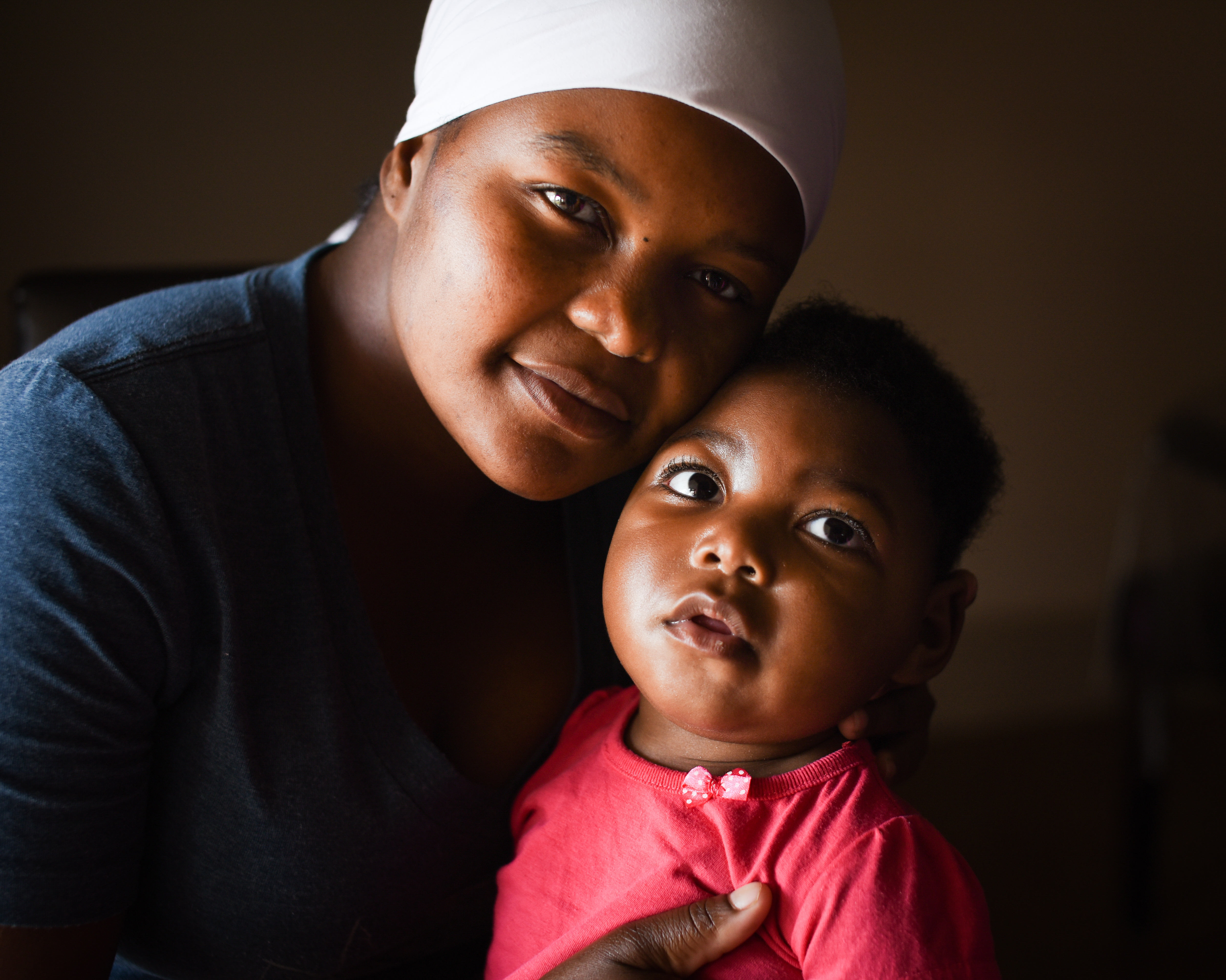
However, when behavior (rather than questionnaire responses) was compared with hormone concentrations, a different story emerged. Blood plasma concentrations of cortisol were positively associated with approach behaviors. In other words, women who had high concentrations of blood cortisol (in samples obtained immediately before or after nursing) engaged in more physically affectionate behaviors (Figure \(\PageIndex{6}\)) and talked more often to their babies than mothers with low cortisol concentrations. Additional analyses from this study revealed that the correlation was even greater for mothers who had reported positive maternal regard (feelings and attitudes) during gestation (pregnancy). Indeed, nearly half of the variation in maternal behavior among women could be accounted for by cortisol concentrations and positive maternal attitudes during pregnancy. Presumably, cortisol does not induce maternal behaviors directly, but it may act indirectly by increasing the mother’s general level of arousal, thus increasing her responsiveness to infant-generated cues. New mothers with high cortisol concentrations were also more attracted to their infant’s odors, were superior in identifying their infants, and generally found cues from infants highly appealing (Fleming, Steiner, & Corter, 1997).
Summary
Hormones are inextricably linked to the development and maturation of reproductive anatomy (both prenatally and during puberty), as well as the promotion and maintenance of adult reproductive functions. The effects of steroid sex hormones are typically characterized as organizational versus activational. Organizational effects result in permanent changes that usually occur early in development, such as the prenatal development of primary sex characteristics (differences in male and female bodies that are present at birth). Activational effects are usually temporary and occur throughout life, dependent on specific conditions.
The main categories of sex hormones are androgens (more prevalent in biological males, e.g. testosterone), and estrogens (more prevalent in biological females, e.g. estradiol). All individuals have both androgens and estrogens, but the relative proportion of the hormones present varies, usually correlated with biological sex. Testosterone is secreted by the testes in males and in small amounts by the ovaries in females. A small amount of testosterone is also secreted by the adrenal glands in both sexes. Estrogens are secreted by the ovaries in females and by fat cells in both sexes.
The interaction between hormones and behavior is bidirectional: hormones can influence behavior, and behavior can sometimes influence hormone concentrations. Hormones do not cause behavioral changes, but they influence the components that interact to produce behavior- sensory systems, the central nervous system, and muscles and glands. In this manner, specific stimuli are more likely to elicit certain responses in the appropriate behavioral or social context. For behavioral effects on hormones, in animal models, losing a fight results in decreased blood testosterone levels. Comparable results have been reported in humans.
Research on rats suggests that the motivation to engage in sexual behavior is separate from the ability to engage in sexual behavior. The hypothalamus plays an important role in motivated behaviors, including sex. In humans, testosterone maintains libido (sex drive) in both males and females.
Although the glans of the clitoris and penis are known to be associated with pleasure, extensive regions of the brain and brainstem are also activated when a person experiences pleasure. Erogenous zones are particularly sensitive areas of skin, which vary across individuals.
The hypothalamus is important for sexual functioning, particularly because it controls the pituitary gland. Hormones related to sexual function secreted by the pituitary gland include follicle-stimulating hormone (FSH), luteinizing hormone (LH), and prolactin from the anterior pituitary, and oxytocin from the posterior pituitary. FSH and LH are responsible for ovulation in females and sperm production in males. Prolactin and oxytocin stimulate lactation. Oxytocin causes rhythmic contractions in the uterus and penis (associated with orgasm), as well as uterine contractions during childbirth. Oxytocin is also believed to be involved in maintaining close relationships.
Hormones trigger the onset of maternal behavior in rats. A fast decline of progesterone in late pregnancy, combined with high concentrations of estradiol, induce female rats to behave maternally (overriding the usual fear response of adult rats toward pups). The medial preoptic area is critical for the expression of rat maternal behavior, and the amygdala appears to inhibit the expression of maternal behavior. Progesterone appears to be the primary hormone that induces maternal aggression (in rats) toward animals that venture too close to their litter.
In human studies, no relationship between hormone concentrations and maternal responsiveness (on attitude questionnaires) was found. However, women who had high concentrations of blood cortisol engaged in more physically affectionate behaviors and talked more often to their babies than mothers with low cortisol concentrations. The correlation was even greater for mothers who had reported positive maternal feelings and attitudes during pregnancy.
References
Arnold, A.P. and Breedlove, S.M. (1985). Organizational and activational effects of sex steroids on brain and behavior: A reanalysis. Hormones and Behavior, 19(4), 469-498.
Fleming, A. S., & Gonzalez, A. (2009). Neurobiology of human maternal care. In P. T. Ellison & P. B. Gray (Eds.), Endocrinology of social relationships (pp. 294–318). Cambridge, MA: Harvard University Press.
Fleming, A. S., Steiner, M., & Corter, C. (1997). Cortisol, hedonics, and maternal responsiveness in human mothers. Hormones and Behavior, 32, 85–98.
Heath, R.G. (1972). Pleasure and brain activity in man. The Journal of Nervous and Mental Disease, 154, 3-18. (As cited in Panksepp, 2004, page 397.)LeVay, S. (1991). A difference in hypothalamic structure between heterosexual and homosexual men. Science, 253(5023), 1034-1037. doi: 10.1126/science.1887219
Jones, R.E. & Lopez, K.H. (2006) Human Reproductive Biology, 3rd edition. Academic Press.
Panksepp, J. (2004). Affective neuroscience: The foundations of human and animal emotions; Chapter 12: The varieties of love and lust: Neural control of sexuality. Oxford University Press.
Additional Resources
- Video: Endocrinology Video (Playlist) - This YouTube playlist contains many helpful videos on the biology of hormones, including reproduction and behavior. This would be a helpful resource for students struggling with hormone synthesis, reproduction, regulation of biological functions, and signaling pathways.
- https://www.youtube.com/playlist?list=PLqTetbgey0aemiTfD8QkMsSUq8hQzv-vA
- Video: Paul Zak: Trust, morality - and oxytocin- This Ted talk explores the roles of oxytocin in the body. Paul Zak discusses biological functions of oxytocin, like lactation, as well as potential behavioral functions, like empathy.
- Video: Sex Differentiation- This video discusses gonadal differentiation, including the role of androgens in the development of male features.
- Video: The Teenage Brain Explained- This is a great video explaining the roles of hormones during puberty.
- Web: Society for Behavioral Neuroendocrinology - This website contains resources on current news and research in the field of neuroendocrinology.
- http://sbn.org/home.aspx
Attributions
- Figures:
- Male/Female "date", goo.gl/m25gce, licensed CC0 Public Domain, sourced from NOBA Hormones & Behavior by Nelson
- Midsagittal brain figure, sourced from OpenStax Psychology 2e Sexual Behavior by Spielman et al. (no author or licensing details given)
- WT and TK rat photo by Jason Snyder from Washington, DC, United States, licensed CC BY 2.0 via Wikimedia Commons
- Colors related to brain arterial supply (mostly) removed by Naomi Bahm, figure originally from Brain areas by Frank Gaillard, https://goo.gl/yCKuQ2, CC-BY-SA 3.0, sourced from NOBA Human Sexual Anatomy and Physiology by Lucas & Fox (who added Identifying asterisks to original image)
- Left image: Hypothalamus location by Blausen.com staff (2014). "Medical gallery of Blausen Medical 2014". WikiJournal of Medicine 1 (2). DOI:10.15347/wjm/2014.010. ISSN 2002-4436., licensed CC BY 3.0 via Wikimedia Commons; Right image: "Hypothalamus controls the pituitary gland", image is in the public domain, sourced from Physical changes in Adolescence by Paris et al.
- Left image: Mother cuddling daughter by Maria Grazia Montagnari, https://goo.gl/LY1Tq0, licensed CC BY 2.0, sourced from NOBA Hormones & Behavior by Nelson; Right image: Mother and young child with a disability by AnikaMeyer, CC BY-SA 4.0 via Wikimedia Commons
- Text adapted from:
- Hormones & Behavior by Randy J. Nelson, licensed CC BY-NC-SA 4.0 via Noba Project.
- "Physical Changes in Adolescence" by Paris, Ricardo, Raymond, & Johnson, LibreTexts is licensed under CC BY.
- " Development of Sexual Identity" by Paris, Ricardo, Raymond, & Johnson, LibreTexts is licensed under CC BY.
- 10.3 Sexual Behavior (Psychology 2e) by Rose M. Spielman, William J. Jenkins, and Marilyn D. Lovett, licensed CC BY 4.0 via OpenStax.
- "Introduction to the Reproductive System" by Suzanne Wakim & Mandeep Grewal, LibreTexts is licensed under CC BY-NC.
- Human Sexual Anatomy and Physiology by Don Lucas and Jennifer Fox, licensed CC BY-NC-SA 4.0 via Noba Project.
- Anabolic Steroids section: "Gonadal and Placental Hormones" by Whitney Menefee, Julie Jenks, Chiara Mazzasette, & Kim-Leiloni Nguyen, LibreTexts is licensed under CC BY.
- Drive States by Sudeep Bhatia and George Loewenstein, licensed CC BY-NC-SA 4.0 via Noba Project. NOTE: Although a few sentences of general text were curated from this source (specifically located in the sections Sexual Behavior as a Form of Motivation, and Sex and the Brain), most of the content it contains (related to the drive state of sexual arousal) is based on rodent research that is presented as though it has been verified to be equivalent to human physiology. In fact, the source they site for male sexuality overlapping with areas associated with aggression specifically states "In many mammals, the vigor of male sexuality and male assertiveness (i.e., social dominance) tend to go together...The fact that male sexuality and aggression interact to a substantial extent in subcortical areas of the brain is now a certainty ... The meaning of this interaction for human sexuality remains regrettably pregnant with ambiguities." (Panksepp 2004, page 239)
- Changes: Text (and images) from above eight sources pieced together with some modifications, transitions and additional content added (particularly the sections: Sex and Hormones, Organizational and Activational Effects of Sex Hormones, and parts of Sex Hormones and Maturation) by Naomi I. Gribneau Bahm, PhD., Psychology Professor at Cosumnes River College, Sacramento, CA.
- (particularly the Sex and Hormones section, Organizational and Activational Effects of Sex Hormones section, and part of the Sex Hormones and Maturation section)