Figure \(\PageIndex{1}\): Phospholipid molecules forming a bilayer with their hydrophobic tails and hydrophilic heads.
Organisms are composed of four basic types of molecules that are essential for cell structure and function: proteins, lipids, carbohydrates, and nucleic acids.Proteins are strings of amino acids that are often folded into complex 3-D shapes. The structure of lipids can be described as having a hydrophilic (water-loving) head and a hydrophobic (water-repelling) tail (Figure 3.1). When lipids are chained together, they form more-complex molecules called fats and triglycerides. Carbohydrates are composed of carbon and hydrogen atoms that can be broken down to supply energy for an organism. Lastly, nucleic acids carry genetic information about a living organism.
Figure \(\PageIndex{2}\): Structural components that form double-stranded nucleic acid (DNA) or single-stranded nucleic acid (RNA).
Probably the most familiar nucleic acid is . DNA comprises a sugar phosphate backbone and nucleotidesno post (Figure 3.2). (More details on the physical structure of DNA and what information DNA nucleotides provide will be discussed later.) Anthropologists can analyze sequences of DNA nucleotides and determine how different organisms are related to each other, since they all have their own unique DNA genetic code. In the case of humans, forensic scientists can identify individuals by analyzing 20 different short DNA sequences known as “CODIS Core Loci.” Another nucleic acid is called . One type of RNA molecule is responsible for chaining amino acids together in order to build proteins (Figure 3.3 and Figure 3.4). How RNA synthesizes amino acids into proteins will be reviewed further on in the chapter.
Figure \(\PageIndex{3}\): Chemical elements that characterize an amino acid. C: carbon; N: Nitrogen; O: Oxygen; H: Hydrogen.Figure \(\PageIndex{4}\): Amino acids (20 different types) strung together form a polypeptide chain.
Cells
In 1665, Robert Hooke observed slices of plant cork using a microscope. Hooke noted that the microscopic plant structures he saw resembled cella, meaning “a small room” in Latin. Approximately two centuries later, biologists recognized the cell as being the most fundamental unit of life and that all life is composed of cells. Cellular organisms can be characterized as two main cell types: prokaryotes and eukaryotes.
Figure \(\PageIndex{5}\): A representation of the single-celled body of E. coli bacteria.
Prokaryotes include bacteria and archaea, and they are composed of a single cell. Additionally, their DNA and organelles are not surrounded by individual membranes. Thus, no compartments separate their DNA from the rest of the cell (Figure 3.5). It is well known that some bacteria can cause illness in humans. For instance, Escherichia coli (E. coli) and Salmonella contamination can result in food poisoning symptoms. Pneumonia and strep throat are caused by Streptococcal bacteria. Neisseria gonorrhoeae is a bacterial sexually transmitted disease. Although bacteria are commonly associated with illness, not all bacteria are harmful. For example, researchers are studying the relationship between the microbiome and human health. The bacteria that are part of the healthy human microbiome perform beneficial roles, such as food digestion, boosting the immune system, and even making vitamins (e.g., B12 and K).
Figure \(\PageIndex{6}\): A microscopic view of plant cell membranes.
Archaea, the other type of prokaryotic organism, were once believed to be closely related to bacteria. However, it was determined through genetic analysis that archaea have their own distinct evolutionary lineage so biologists reclassified them into their own taxonomic domain. Archaea were discovered living in extreme environments and are therefore known as “extremophiles.” For example, archaea can be found in high temperatures, such as Old Faithful Geyser in Yellowstone National Park.
Eukaryotes can be single-celled or multicelled in their body composition. In contrast to prokaryotes, eukaryotes possess membranes that surround their DNA and organelles. An example of a single-celled eukaryote is the microscopic algae found in ponds (phytoplankton), which can produce oxygen from the sun. Yeasts are also single-celled, and fungi can be single- or multicellular. Plants and animals are all multicellular.
Although plant and animal cells have a surprising number of similarities, there are some key differences. For example, plant cells possess a thick outer cell membrane made of a fibrous carbohydrate called cellulose (Figure 3.6). Animal and plant cells also have different tissues. A tissue is an aggregation of cells that are morphologically similar and perform the same task. For most plants, the outermost layer of cells forms a waxy cuticle that helps to protect the cells and to prevent water loss. However, humans have skin, the outermost cell layer of which is mostly composed of a tough protein called keratin. Overall, humans have a diversity of tissue types (e.g., cartilage, brain, and heart).
Animal Cell Organelles
Figure \(\PageIndex{7}\): A phospholipid bilayer with membrane-bound carbohydrates and proteins.
An animal cell is surrounded by a double membrane called the phospholipid bilayer (Figure 3.7). A closer look reveals that this protective barrier is made of lipids and proteins that provide structure and function for cellular activities. For example, lipids and proteins embedded in the cell’s membrane work together to regulate the passage of molecules and ions (e.g., H2O and sodium) into and out of the cell. Cytoplasm is the jelly-like matrix inside of the cell membrane. Part of the cytoplasm comprises organelles, which perform different specialized tasks for the cell (Figure 3.8). An example of an organelle is the nucleus, where the cell’s DNA is located (Figure 3.9). The double membrane that encloses the nucleus is known as the nuclear envelope; its purpose is to regulate molecules into and out of the nucleus and serve as a barrier to protect DNA integrity.
Figure \(\PageIndex{8}\): An animal cell with membrane-enclosed organelles.Figure \(\PageIndex{9}\): A membrane-enclosed nucleus of an animal cell.
Another important organelle is the mitochondrion (Figure 3.10). Mitochondria are often referred to as “powerhouse centers” because they produce energy for the cell in the form of adenosine triphosphate (ATP)no post. Depending on the species and tissue type, multicellular eukaryotes can have hundreds to thousands of mitochondria in each of their cells. Scientists have determined that mitochondria played an important role in the evolution of the eukaryotic cell. Mitochondria were once symbiotic prokaryotic organisms (i.e., helpful bacteria) that transformed into cellular organelles over time. Because mitochondria used to be separate organisms, this explains why mitochondria also have their own DNA, called . All organelles have important physiological functions, and when they cannot perform their role optimally, it can result in disease. For example, there are mitochondrial diseases for which cells have abnormally less mitochondria. In humans, this leads to various neurological symptoms and disorders. Figure 3.11 lists other organelles found in the cell and their specialized cellular roles.
Figure \(\PageIndex{10}\): Microscopic view of an animal mitochondrion organelle.
Table 3.1.1: Names of organelles and their cellular functions.
Cell structure
Description
Cytoplasm
Fluid substance located inside of cell membrane that contains organelles
Nucleopore
Pores in the nuclear envelope that are selectively permeable
Nucleus
Contains the cell’s DNA and is surrounded by the nuclear envelope
Nucleolus
Resides inside of the nucleus and is the site of ribosomal RNA (rRNA) transcription, processing, and assembly
Mitochondrion
Responsible for cellular respiration, where energy is produced by converting nutrients into ATP
Ribosome
Located in the cytoplasm and also the membrane of the rough endoplasmic reticulum. Messenger RNA (mRNA) binds to ribosomes and proteins are synthesized
Endoplasmic reticulum (ER)
Continuous membrane with the nucleus that helps transport, synthesize, modify, and fold proteins. Rough ER has embedded ribosomes, whereas smooth ER lacks ribosomes
Golgi body
Layers of flattened sacs that receive and transmit messages from the ER to secrete and transport proteins within the cell
Lysosome
Located in the cytoplasm and contains enzymes to degrade cellular components
Microtubule
Involved with cellular movement including intracellular transport and cell division
Centrioles
Assist with the organization of mitotic spindles which extend and contract for the purpose of cellular movement during mitosis and meiosis

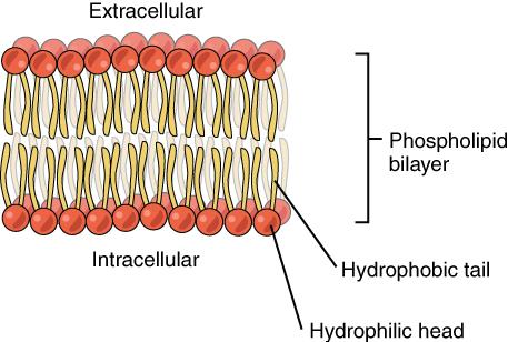 Figure \(\PageIndex{1}\): Phospholipid molecules forming a bilayer with their hydrophobic tails and hydrophilic heads.
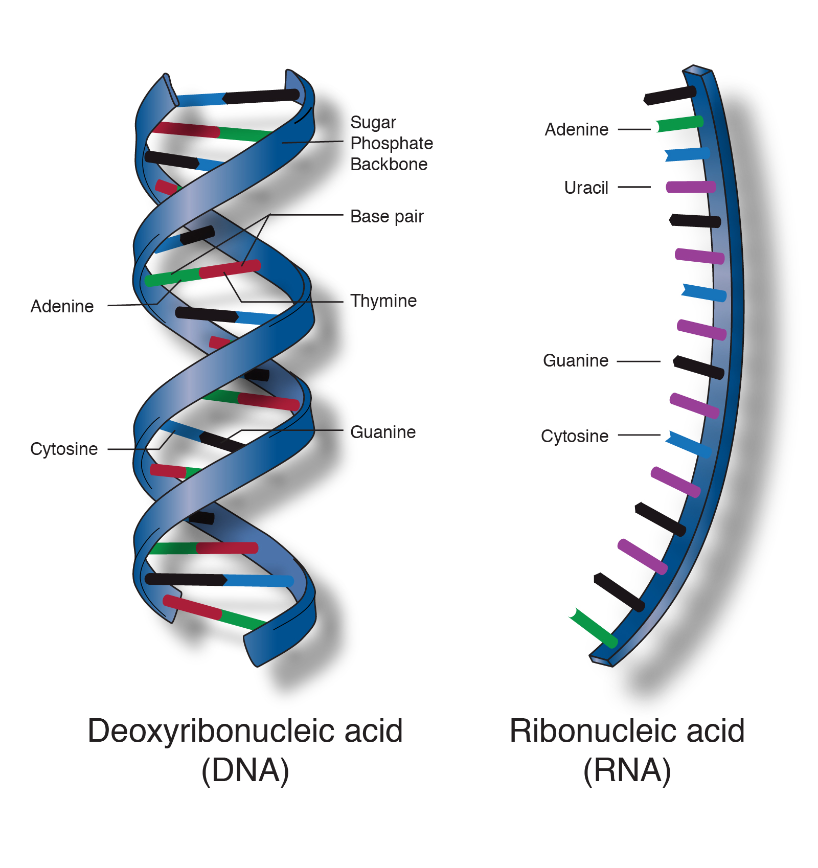 Figure \(\PageIndex{2}\): Structural components that form double-stranded nucleic acid (DNA) or single-stranded nucleic acid (RNA).
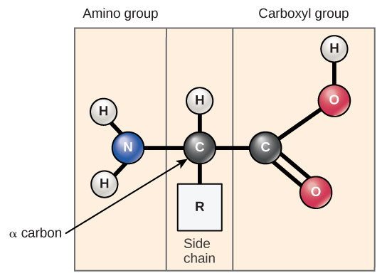 Figure \(\PageIndex{3}\): Chemical elements that characterize an amino acid. C: carbon; N: Nitrogen; O: Oxygen; H: Hydrogen.
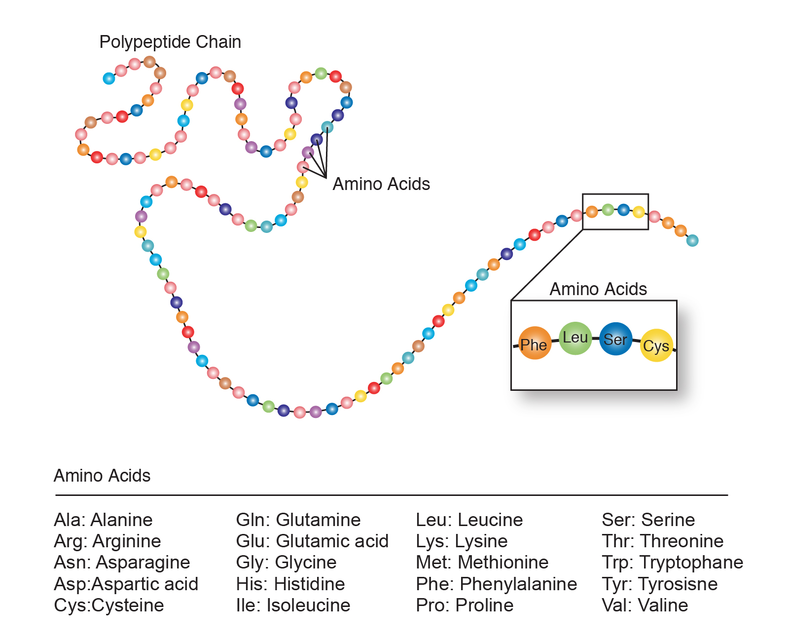 Figure \(\PageIndex{4}\): Amino acids (20 different types) strung together form a polypeptide chain.
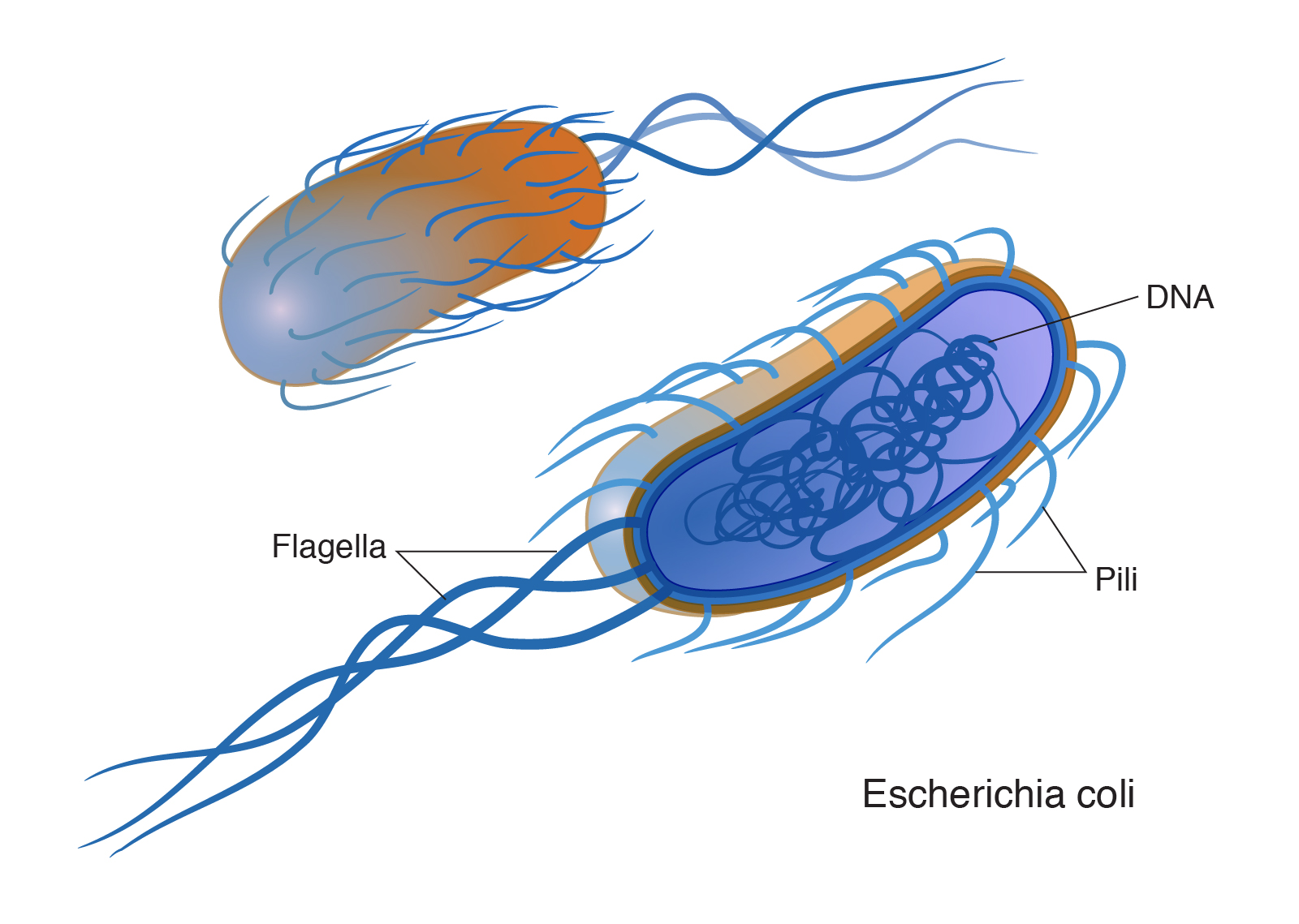 Figure \(\PageIndex{5}\): A representation of the single-celled body of E. coli bacteria.
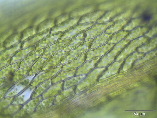 Figure \(\PageIndex{6}\): A microscopic view of plant cell membranes.
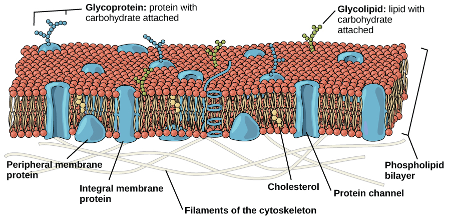 Figure \(\PageIndex{7}\): A phospholipid bilayer with membrane-bound carbohydrates and proteins.
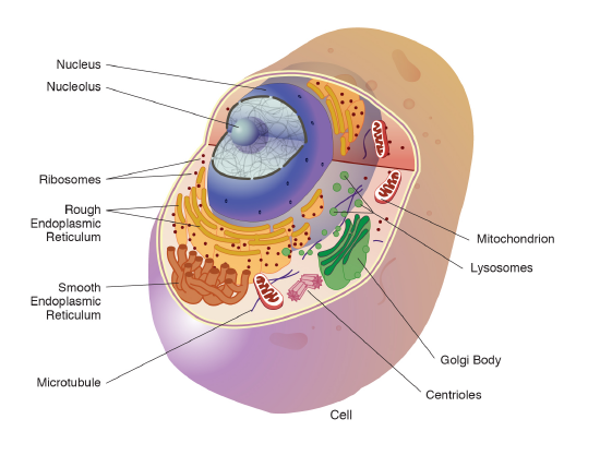 Figure \(\PageIndex{8}\): An animal cell with membrane-enclosed organelles.
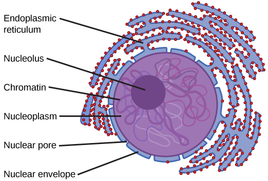 Figure \(\PageIndex{9}\): A membrane-enclosed nucleus of an animal cell.
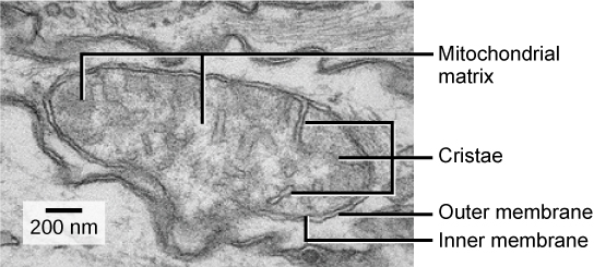 Figure \(\PageIndex{10}\): Microscopic view of an animal mitochondrion organelle.