

15.4: Trauma Analysis and Bone Pathology
- Page ID
- 158821
TYPES OF TRAUMA
An injury to living tissue caused by an extrinsic force or mechanism. (See Lovell 1997, 139.)
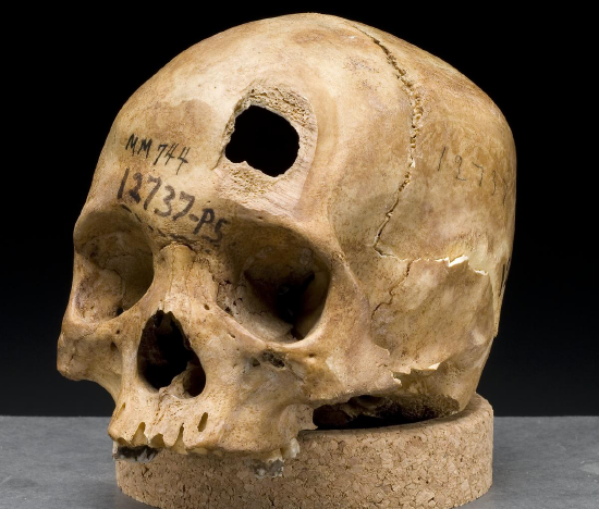 Figure \(\PageIndex{1}\): Example of sharp-force trauma (sword wound) to the frontal bone.
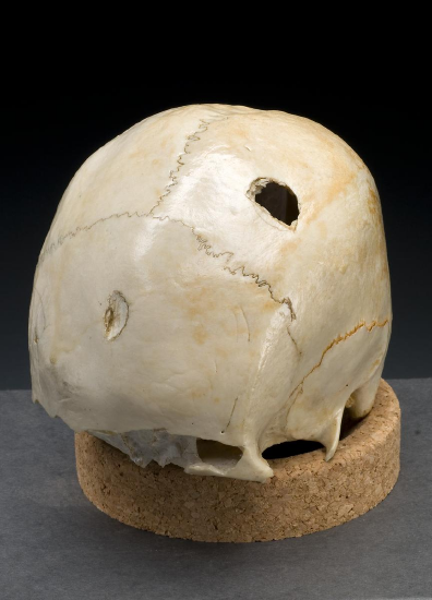 Figure \(\PageIndex{2}\): Example of multiple blunt force impacts to the left parietal and frontal bones.
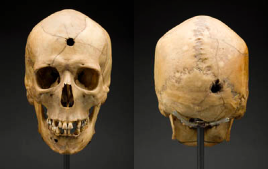 Figure \(\PageIndex{3}\): Example of projectile trauma with an entrance wound to the frontal bone and exit wound visible on the occipital.
Timing of Injury
Trauma occurring before death.
Trauma occurring at or around the time of death.
Trauma occurring after death.
The Role of the Forensic Anthropologist in Trauma Analysis
While there is a wide range of variation within the human skeletal system, bone development can also occur pathologically. Bone pathology can occur when there is excessive bone growth (osteoblastic activity or bone building) or bone is destroyed unnecessarily (osteoclastic activity or bone breakdown). Osteoblastic (bone building) and osteoclastic (bone destruction or breakdown) activities are normal processes of bone development, growth, and maintenance; however, when bone growth or breakdown exceeds what is necessary, the bony change can be classified as pathological, resulting in a bone pathology.
TYPES OF BONE PATHOLOGY
For the purposes of this chapter, we will focus on both osteoblastic and osteoclastic pathologies of the human skeleton. In addition to considering whether a pathology is osteoclastic or osteoblastic, it is also important to classify a pathology according to its origin. Bone pathologies can be classified in a number of ways, including:
- congenital: occurring in the developmental period, often hereditary;
- traumatic: resulting from extrinsic factors and forces;
- degenerative: causing the degeneration or breakdown of bone tissue;
- infectious: resulting from bacterial, viral, or fungal agents;
- circulatory: resulting from a disruption in the relationship between the skeletal and circulatory system;
- metabolic: resulting from nutrient deficiencies;
- endocrinological: caused by hormonal imbalances; and
- neoplastic: related to abnormal growth, both benign and malignant, of bone tissue.
For the remainder of this section, we will focus on six different bone pathologies: (1) osteosarcoma, (2) osteogenesis imperfecta, (3) rickets, (4) achondroplasia, (5) Paget’s disease of bone, and (6) diffuse idiopathic skeletal hyperostosis (DISH).
Osteosarcoma
Osteosarcoma is a type of neoplastic bone pathology. Characterized by malignant tumors that begin within bone tissues, osteosarcoma is a primary bone cancer (meaning it begins directly in bone tissue, rather than spreading to bone from other body tissues). Malignant tumors associated with osteosarcoma usually occur during growth and development and are observed most often in adolescents and young adults (Ortner and Putschar 1981, 384). Tumors are most frequently observed near the ends of long bones (Figure \(\PageIndex{4}\); Ortner and Putschar 1981, 384).
 Figure \(\PageIndex{4}\) Osteosarcoma on a left human femur.
Osteogenesis Imperfecta
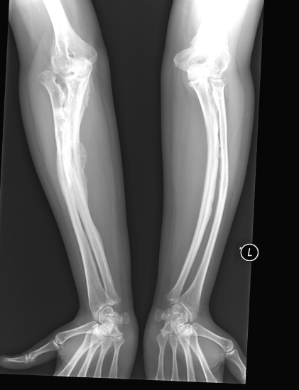 Figure \(\PageIndex{5}\): X-ray of the forearms of an individual with osteogenesis imperfecta (note the presence of multiple healing fractures).
Osteogenesis Imperfecta (OI) is a congenital bone pathology characterized by bones with low collagen content, leading to frequent fracturing (Ortner and Putschar 1981, 337). However, OI can also occur as a result of a spontaneous mutation. The disease is characterized by multiple fractures throughout the skeleton, particularly in the long bones (Figure \(\PageIndex{5}\)). Depending on the type of OI, the disease is either manifest at birth or during childhood or adolescence (Ortner and Putschar 1981, 337). In addition to their susceptibility to easily fractured bones, individuals with OI are typically shorter in stature and may be subject to fracturing of tooth enamel and premature tooth loss (Ortner and Putschar 1981, 337).
Rickets
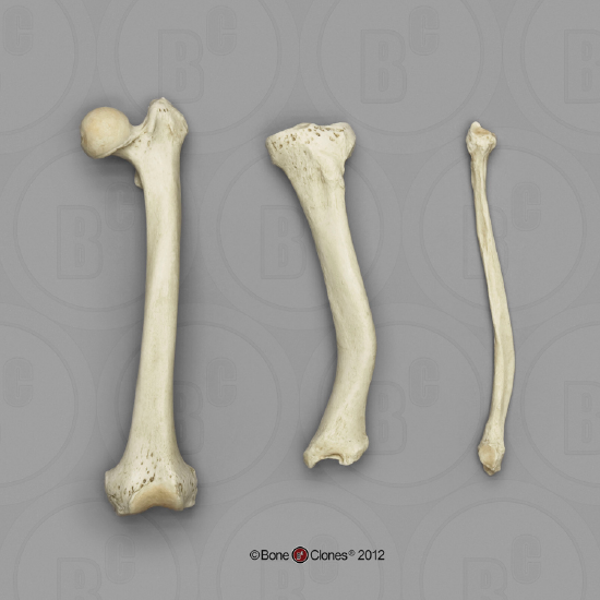 Figure \(\PageIndex{6}\): Example of rickets in long bones of the leg.
Rickets is a metabolic bone pathology resulting from a Vitamin D deficiency in childhood (Ortner and Putschar 1982, 273). Vitamin D is essential to the mineralization of bone tissue and is characterized by a wide variety of cranial and postcranial changes, including the following: asymmetrical deformities of the skull, bowing of the long bones, vertebral compression fractures, and a smaller, thicker pelvis (Figure \(\PageIndex{6}\); Ortner and Putschar 1981, 273–278).
Achondroplasia
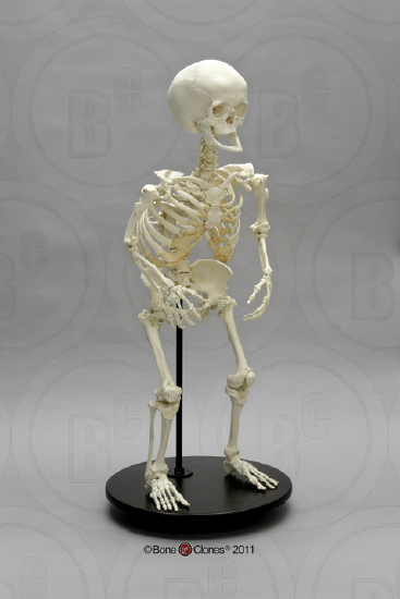 Figure \(\PageIndex{7}\): A cast of a complete skeleton of an adult female skeleton with achondroplasia.
Achondroplasia is a congenital bone pathology resulting from an abnormality in the conversion of cartilage to bone and is the most common form of dwarfism (Ortner and Putschar 1981, 329). The skeletal manifestations of achondroplasia are most apparent in the long bones comprising the arms and legs, while the trunk is of relatively normal proportions in individuals with achondroplasia (Figure \(\PageIndex{7}\)). On average, males with achondroplasia are approximately 4'4" tall and females are approximately 4'1" tall (NIH 2019).
Paget’s Disease of Bone
Paget’s disease of bone is a disease of unknown origin that causes bones to grow larger and weaker over time (NIH 2019). The disease is marked by both osteoblastic and osteoclastic activity, with excessive osteoclastic resorption followed by osteoblastic proliferation leading to unnecessary amounts of new woven bone (Ortner and Putschar 1981, 309). The disease typically does not appear until the fourth or fifth decade of life and is more common in males than females (Ortner and Putschar 1981, 309). Paget’s disease of bone can affect any bone, but the most commonly affected elements include the spine, pelvis, skull, and legs. The frequency of osteosarcoma is also higher among individuals with Paget’s disease of bone (NIH 2019).
Diffuse Idiopathic Skeletal Hyperostosis (DISH)
DISH is a bone pathology characterized by a hardening (calcification or buildup of calcium salts) of the ligaments and tendons of the vertebral column. While DISH is observed in other areas of the skeleton, the vertebral column is the most frequently affected region. DISH is more prevalent in males than females and typically is observed in older adults (50-plus years) (NIH 2019). Recent medical research suggests that DISH results from abnormal osteoblastic activity in the spine, leading to excessive bone growth (NIH 2019).
REFERENCES
Galloway, Alison. Broken Bones: Anthropological Analysis of Blunt Force Trauma. 1999. Springfield, IL: Charles C. THomas Publisher, LTD.
Lovell, Nancy C. 1997. “Trauma Analysis in Paleopathology.” Yearbook of Physical Anthropology 104 (S25): 139–170.
NIH U.S. National Library of Medicine. 2019. “Genetics Home Reference: Achondroplasia.” Last modified February 5, 2019. https://ghr.nlm.nih.gov/condition/achondroplasia.
Ortner, Donald J., and Walter G. J. Putschar. 1981. Identification of Pathological Conditions in Human Skeletal Remains. Washington, D.C.: Smithsonian Institution Press.
Scientific Working Group for Forensic Anthropology (SWGANTH). 2010b. “Sex Assessment.” Last modified June 3, 2010. www.nist.gov/sites/default/files/documents/2018/03/13/swganth_sex_assessment.pdf.
Scientific Working Group for Forensic Anthropology (SWGANTH). 2011. “Trauma Analysis.” Last modified May 27, 2011. www.nist.gov/sites/default/files/documents/2018/03/13/swganth_trauma.pdf.
FIGURE ATTRIBUTIONS
Figure \(\PageIndex{1}\) Skull sword trauma by the National Institutes of Health, Health & Human Services [19th Century Collection, National Museum of Health and Medicine, Armed Forces Institute of Pathology, Washington, D.C. From exhibition “Visible Proofs: Forensic Views of the Body” U.S. National Library of Medicine] is in the public domain.
Figure \(\PageIndex{2}\) Skull hammer trauma by the National Institutes of Health, Health & Human Services [19th Century Collection, National Museum of Health and Medicine, Armed Forces Institute of Pathology, Washington, D.C. From exhibition “Visible Proofs: Forensic Views of the Body” U.S. National Library of Medicine] is in the public domain.
Figure \(\PageIndex{3}\) Trauma: Gunshot Wounds by Smithsonian [exhibit: Written in Bone, How Bone Biographies Get Written] has no known copyright restrictions.
Figure \(\PageIndex{4}\) Human Left Femur, Osteosarcoma by ©BoneClones is used by permission and available here is under a CC BY-NC 4.0 License.
Figure \(\PageIndex{5}\) Osteogenesis Imperfecta Type V by ShakataGaNai is used under a CC BY-SA 4.0 License.
Figure \(\PageIndex{6}\) Human Femur, Tibia and Fibula, Rickets by ©BoneClones is used by permission and available here is under a CC BY-NC 4.0 License.
Figure \(\PageIndex{7}\) Human Female Achondroplasia Dwarf Skeleton, Articulated by ©BoneClones is used by permission and available here is under a CC BY-NC 4.0 License.