
2.1: Synapse Structure
- Page ID
- 170807
\( \newcommand{\vecs}[1]{\overset { \scriptstyle \rightharpoonup} {\mathbf{#1}} } \)
\( \newcommand{\vecd}[1]{\overset{-\!-\!\rightharpoonup}{\vphantom{a}\smash {#1}}} \)
\( \newcommand{\id}{\mathrm{id}}\) \( \newcommand{\Span}{\mathrm{span}}\)
( \newcommand{\kernel}{\mathrm{null}\,}\) \( \newcommand{\range}{\mathrm{range}\,}\)
\( \newcommand{\RealPart}{\mathrm{Re}}\) \( \newcommand{\ImaginaryPart}{\mathrm{Im}}\)
\( \newcommand{\Argument}{\mathrm{Arg}}\) \( \newcommand{\norm}[1]{\| #1 \|}\)
\( \newcommand{\inner}[2]{\langle #1, #2 \rangle}\)
\( \newcommand{\Span}{\mathrm{span}}\)
\( \newcommand{\id}{\mathrm{id}}\)
\( \newcommand{\Span}{\mathrm{span}}\)
\( \newcommand{\kernel}{\mathrm{null}\,}\)
\( \newcommand{\range}{\mathrm{range}\,}\)
\( \newcommand{\RealPart}{\mathrm{Re}}\)
\( \newcommand{\ImaginaryPart}{\mathrm{Im}}\)
\( \newcommand{\Argument}{\mathrm{Arg}}\)
\( \newcommand{\norm}[1]{\| #1 \|}\)
\( \newcommand{\inner}[2]{\langle #1, #2 \rangle}\)
\( \newcommand{\Span}{\mathrm{span}}\) \( \newcommand{\AA}{\unicode[.8,0]{x212B}}\)
\( \newcommand{\vectorA}[1]{\vec{#1}} % arrow\)
\( \newcommand{\vectorAt}[1]{\vec{\text{#1}}} % arrow\)
\( \newcommand{\vectorB}[1]{\overset { \scriptstyle \rightharpoonup} {\mathbf{#1}} } \)
\( \newcommand{\vectorC}[1]{\textbf{#1}} \)
\( \newcommand{\vectorD}[1]{\overrightarrow{#1}} \)
\( \newcommand{\vectorDt}[1]{\overrightarrow{\text{#1}}} \)
\( \newcommand{\vectE}[1]{\overset{-\!-\!\rightharpoonup}{\vphantom{a}\smash{\mathbf {#1}}}} \)
\( \newcommand{\vecs}[1]{\overset { \scriptstyle \rightharpoonup} {\mathbf{#1}} } \)
\( \newcommand{\vecd}[1]{\overset{-\!-\!\rightharpoonup}{\vphantom{a}\smash {#1}}} \)
For the nervous system to function, neurons must be able to communicate with each other, and they do this through structures called synapses. At the synapse, the terminal of a presynaptic cell comes into close contact with the cell membrane of a postsynaptic neuron.

Synapse Types
There are two types of synapses: electrical and chemical.
Electrical
Electrical synapses are a direct connection between two neurons. Cell membrane proteins called connexons form gap junctions between the neurons. The gap junctions form pores that allow ions to flow between neurons, so as an action potential propagates in the presynaptic neuron, the influx of sodium can move directly into the postsynaptic neuron and depolarize the cell. The response in the postsynaptic cell is almost immediate, with little to no delay between signaling in the pre- and postsynaptic neurons.


Animation 8.1. Membrane-bound proteins called connexons form gap junctions between presynaptic and postsynaptic neurons. This allows for direct exchange of ions between neurons. An action potential in the presynaptic neuron will cause an immediate depolarization of the postsynaptic membrane because the sodium ions will cross the membrane through the gap junctions. ‘Electrical Synapse – Ion Flow’ by Casey Henley is licensed under a Creative Commons Attribution Non-Commercial Share-Alike (CC BY-NC-SA) 4.0 International License. View static image of animation.
Since the gap junctions allow diffusion of ions without any obstruction, the signal can flow bidirectionally through an electrical synapse. The electrochemical gradients will drive direction of ion flow.
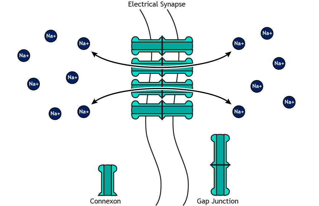
Animation 8.2. Since an electrical synapse is a direct, physical connection between two neurons, ions are able to flow either direction across the gap junction. ‘Bidirectional Electrical Synapse’ by Casey Henley is licensed under a Creative Commons Attribution Non-Commercial Share-Alike (CC BY-NC-SA) 4.0 International License. View static image of animation.
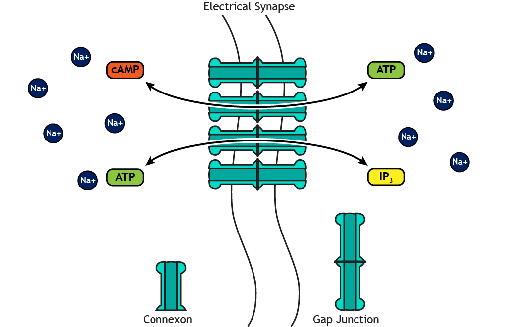
Additionally, small molecules like ATP or second messengers can also move through the gap junctions. These signaling molecules play an important role in cellular mechanisms, which we will see in a later chapter.
Animation 8.3. Gap junctions are large enough to allow the flow of small cellular molecules like ATP or second messengers. ‘Electrical Synapse – Small Molecules’ by Casey Henley is licensed under a Creative Commons Attribution Non-Commercial Share-Alike (CC BY-NC-SA) 4.0 International License. View static image of animation.
Chemical
Chemical synapses do not form physical connections between the pre- and postsynaptic neurons. Instead, a space called the synaptic cleft exists between the presynaptic terminal and the postsynaptic membrane.

Figure 8.2. A chemical synapse does not make direct contact between the two neurons. The presynaptic terminal and the postsynaptic membrane are separated by the synaptic cleft. Neurotransmitters are stored in the presynaptic cell, and the postsynaptic cell has neurotransmitter receptors in the membrane. ‘Chemical Synapse’ by Casey Henley is licensed under a Creative Commons Attribution Non-Commercial Share-Alike (CC BY-NC-SA) 4.0 International License.
At a chemical synapse, the depolarization of an action potential reaching the presynaptic terminal causes release of neurotransmitters, which act on specialized receptors located in the cell membrane of the postsynaptic neuron. The structure and function of chemical synapses make them slower than electrical synapses and permit signaling in only one direction.
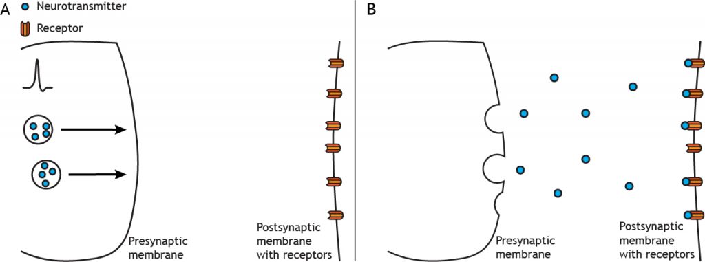
Animation 8.4. An action potential causes release of neurotransmitters from the presynaptic terminal into the synaptic cleft. The transmitters then act on neurotransmitter receptors in the postsynaptic membrane. ‘Chemical Synapse – Neurotransmitter Release’ by Casey Henley is licensed under a Creative Commons Attribution Non-Commercial Share-Alike (CC BY-NC-SA) 4.0 International License. View static image of animation.
Synapse Location
As we discuss synaptic transmission, we will focus mainly on axodendritic synapses, in which the presynaptic terminal synapses on the dendrites of the postsynaptic cell. But synapses can also be located between the terminal and the cell body of the postsynaptic cell, called axosomatic, or even between the terminal and the axon of the postsynaptic cell, called axoaxonic.

Key Takeaways
- Electrical synapses make direct contact between neurons, are faster than chemical synapses, and can be bidirectional
- Chemical synapses form a synaptic cleft between the neurons and are unidirectional
- Synapses can occur between the presynaptic terminal and the postsynaptic dendrites (axodendritic), cell body (axosomatic), or axon (axoaxonic)