
5.1: Neurons and their Basic Functions
- Page ID
- 173126
\( \newcommand{\vecs}[1]{\overset { \scriptstyle \rightharpoonup} {\mathbf{#1}} } \)
\( \newcommand{\vecd}[1]{\overset{-\!-\!\rightharpoonup}{\vphantom{a}\smash {#1}}} \)
\( \newcommand{\id}{\mathrm{id}}\) \( \newcommand{\Span}{\mathrm{span}}\)
( \newcommand{\kernel}{\mathrm{null}\,}\) \( \newcommand{\range}{\mathrm{range}\,}\)
\( \newcommand{\RealPart}{\mathrm{Re}}\) \( \newcommand{\ImaginaryPart}{\mathrm{Im}}\)
\( \newcommand{\Argument}{\mathrm{Arg}}\) \( \newcommand{\norm}[1]{\| #1 \|}\)
\( \newcommand{\inner}[2]{\langle #1, #2 \rangle}\)
\( \newcommand{\Span}{\mathrm{span}}\)
\( \newcommand{\id}{\mathrm{id}}\)
\( \newcommand{\Span}{\mathrm{span}}\)
\( \newcommand{\kernel}{\mathrm{null}\,}\)
\( \newcommand{\range}{\mathrm{range}\,}\)
\( \newcommand{\RealPart}{\mathrm{Re}}\)
\( \newcommand{\ImaginaryPart}{\mathrm{Im}}\)
\( \newcommand{\Argument}{\mathrm{Arg}}\)
\( \newcommand{\norm}[1]{\| #1 \|}\)
\( \newcommand{\inner}[2]{\langle #1, #2 \rangle}\)
\( \newcommand{\Span}{\mathrm{span}}\) \( \newcommand{\AA}{\unicode[.8,0]{x212B}}\)
\( \newcommand{\vectorA}[1]{\vec{#1}} % arrow\)
\( \newcommand{\vectorAt}[1]{\vec{\text{#1}}} % arrow\)
\( \newcommand{\vectorB}[1]{\overset { \scriptstyle \rightharpoonup} {\mathbf{#1}} } \)
\( \newcommand{\vectorC}[1]{\textbf{#1}} \)
\( \newcommand{\vectorD}[1]{\overrightarrow{#1}} \)
\( \newcommand{\vectorDt}[1]{\overrightarrow{\text{#1}}} \)
\( \newcommand{\vectE}[1]{\overset{-\!-\!\rightharpoonup}{\vphantom{a}\smash{\mathbf {#1}}}} \)
\( \newcommand{\vecs}[1]{\overset { \scriptstyle \rightharpoonup} {\mathbf{#1}} } \)
\( \newcommand{\vecd}[1]{\overset{-\!-\!\rightharpoonup}{\vphantom{a}\smash {#1}}} \)
\(\newcommand{\avec}{\mathbf a}\) \(\newcommand{\bvec}{\mathbf b}\) \(\newcommand{\cvec}{\mathbf c}\) \(\newcommand{\dvec}{\mathbf d}\) \(\newcommand{\dtil}{\widetilde{\mathbf d}}\) \(\newcommand{\evec}{\mathbf e}\) \(\newcommand{\fvec}{\mathbf f}\) \(\newcommand{\nvec}{\mathbf n}\) \(\newcommand{\pvec}{\mathbf p}\) \(\newcommand{\qvec}{\mathbf q}\) \(\newcommand{\svec}{\mathbf s}\) \(\newcommand{\tvec}{\mathbf t}\) \(\newcommand{\uvec}{\mathbf u}\) \(\newcommand{\vvec}{\mathbf v}\) \(\newcommand{\wvec}{\mathbf w}\) \(\newcommand{\xvec}{\mathbf x}\) \(\newcommand{\yvec}{\mathbf y}\) \(\newcommand{\zvec}{\mathbf z}\) \(\newcommand{\rvec}{\mathbf r}\) \(\newcommand{\mvec}{\mathbf m}\) \(\newcommand{\zerovec}{\mathbf 0}\) \(\newcommand{\onevec}{\mathbf 1}\) \(\newcommand{\real}{\mathbb R}\) \(\newcommand{\twovec}[2]{\left[\begin{array}{r}#1 \\ #2 \end{array}\right]}\) \(\newcommand{\ctwovec}[2]{\left[\begin{array}{c}#1 \\ #2 \end{array}\right]}\) \(\newcommand{\threevec}[3]{\left[\begin{array}{r}#1 \\ #2 \\ #3 \end{array}\right]}\) \(\newcommand{\cthreevec}[3]{\left[\begin{array}{c}#1 \\ #2 \\ #3 \end{array}\right]}\) \(\newcommand{\fourvec}[4]{\left[\begin{array}{r}#1 \\ #2 \\ #3 \\ #4 \end{array}\right]}\) \(\newcommand{\cfourvec}[4]{\left[\begin{array}{c}#1 \\ #2 \\ #3 \\ #4 \end{array}\right]}\) \(\newcommand{\fivevec}[5]{\left[\begin{array}{r}#1 \\ #2 \\ #3 \\ #4 \\ #5 \\ \end{array}\right]}\) \(\newcommand{\cfivevec}[5]{\left[\begin{array}{c}#1 \\ #2 \\ #3 \\ #4 \\ #5 \\ \end{array}\right]}\) \(\newcommand{\mattwo}[4]{\left[\begin{array}{rr}#1 \amp #2 \\ #3 \amp #4 \\ \end{array}\right]}\) \(\newcommand{\laspan}[1]{\text{Span}\{#1\}}\) \(\newcommand{\bcal}{\cal B}\) \(\newcommand{\ccal}{\cal C}\) \(\newcommand{\scal}{\cal S}\) \(\newcommand{\wcal}{\cal W}\) \(\newcommand{\ecal}{\cal E}\) \(\newcommand{\coords}[2]{\left\{#1\right\}_{#2}}\) \(\newcommand{\gray}[1]{\color{gray}{#1}}\) \(\newcommand{\lgray}[1]{\color{lightgray}{#1}}\) \(\newcommand{\rank}{\operatorname{rank}}\) \(\newcommand{\row}{\text{Row}}\) \(\newcommand{\col}{\text{Col}}\) \(\renewcommand{\row}{\text{Row}}\) \(\newcommand{\nul}{\text{Nul}}\) \(\newcommand{\var}{\text{Var}}\) \(\newcommand{\corr}{\text{corr}}\) \(\newcommand{\len}[1]{\left|#1\right|}\) \(\newcommand{\bbar}{\overline{\bvec}}\) \(\newcommand{\bhat}{\widehat{\bvec}}\) \(\newcommand{\bperp}{\bvec^\perp}\) \(\newcommand{\xhat}{\widehat{\xvec}}\) \(\newcommand{\vhat}{\widehat{\vvec}}\) \(\newcommand{\uhat}{\widehat{\uvec}}\) \(\newcommand{\what}{\widehat{\wvec}}\) \(\newcommand{\Sighat}{\widehat{\Sigma}}\) \(\newcommand{\lt}{<}\) \(\newcommand{\gt}{>}\) \(\newcommand{\amp}{&}\) \(\definecolor{fillinmathshade}{gray}{0.9}\)Learning Objectives
- Describe the anatomy of neurons and the function of each of the three major parts of a neuron
- Describe neurotransmitters, synapse, synaptic vesicles, resting potential, EPSP, IPSP, and action potential
- Explain saltatory conduction and why it is important to communication within the nervous system; include description of the myelin sheath and nodes of Ranvier
- Describe the role of post-synaptic receptor sites in communication between neurons
- Describe neurons classified by shape and by their functions
Overview
In this section, we continue our exploration of neurons, the building blocks of the nervous system. We examine how they generate electrochemical signals, and how the billions of neurons in the nervous system communicate with one another, a process known as synaptic transmission. Before tackling these topics, we review and expand the basic anatomy and functioning of neurons covered in part in Chapter 4. A sound grasp of these facts provides the groundwork for understanding how neuron potentials are generated within neurons and how they combine to trigger synaptic transmission. As you read, remember that the voltages and chemical events we discuss in this section, operating in large populations of brain cells, somehow generate our perceptions, thoughts, emotions, and the entirety of our mental experience. To date, how this happens, how patterns of neuron potentials in brain circuits become conscious minds, remains the greatest mystery of all facing modern science.
Neuron Anatomy and The Synapse
To understand neuron function, it is important to review the neuronal anatomy presented previously.
Although there are some exceptions, neurons have three major structural parts - - the soma or cell body, dendrites (the "receivers" of the neuron), and the axon (the ouput end of the neuron). The entire neuron is bounded by a cell membrane, the neuronal membrane. The neuronal membrane of a neuron has channels or "doors" for ions (electrically charged atoms) which are able to pass through the membrane when specific channels are opened. Figure \(\PageIndex{1}\) shows this basic neuron anatomy.
The soma or cell body contains organelles, such as ribosomes and mitochondria which are common to most types of cells in the body. These are involved in the basic metabolism of the cell. The soma also contains the nucleus, where the genes and chromosomes (containing DNA) are located.
The second main part of the neuron are the dendrites, the information receivers of the neuron. Dendrites in some neurons can branch profusely (dendritic trees), expanding the region of the neuron that can receive inputs from other neurons. The receptor sites (or more technically, postsynaptic receptor sites because of their location on the receiving or postsynaptic neuron) which receive molecules of neurotransmitter are located on the dendrites (and, to a lesser degree, on the soma) of the receiving neuron. On the dendrites are small dendritic spines which are associated with the connections between neurons (the synapses) and can change shape rapidly when learning occurs. Notice that the spines are not the same thing as dendritic branches. In Figure \(\PageIndex{2}\), a dendritic branch with dendritic spines is shown on the left in microscopic detail, and on the right, are dendritic trees of two types of neuron found in the retina. Spines are not visible in the images of dendritic trees on the right because the dendritic spines are too small, while dendritic branches comprising the dendritic trees can easily be seen (see caption for Figure \(\PageIndex{2}\)).
Note: A detailed discussion of the structure of the synapse and synaptic communication will be covered later in this chapter.
.jpg?revision=1)
The third major part of the neuron is the axon, coming out of the soma like a hose. The axon carries the output messages of a neuron (nerve impulses) along its length to its axon terminals (axon endings). There is only one axon per neuron, although it can branch into multiple axon terminals. In a typical neuron, the root end of the axon emerges out of the soma at a small swelling called the axon hillock. Between the axon hillock and the first segment of the axon is where the nerve impulse is first generated (see discussion of the action potential, the nerve impulse, that follows below).
Myelinated and Unmyelinated Axons
Some axons have a glial cell covering known as the myelin sheath. The glial cells capable of producing myelin include oligodendrocytes of the CNS and the Schwann cells of the PNS. This fatty, insulating, myelin sheath has gaps in it, revealing the bare axon, at regular intervals along the axon's length. These bare spots along the length of a myelinated axon are called nodes, or nodes of Ranvier, after their discoverer.
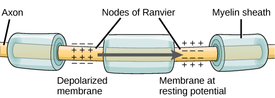
Figure \(\PageIndex{3}\): Nodes of Ranvier. Nodes of Ranvier are gaps in the myelin sheath which covers myelinated axons. Nodes contain voltage-gated potassium (K+) and sodium (Na+) channels. Action potentials travel down the axon by "jumping"from one node to the next, speeding conduction of the action potential down the length of the axon toward the axon ending, also known as the axon bouton, axon button, or axon terminal.
The function of the myelin sheath and the nodes is to speed up the rate at which nerve impulses travel down the length of the axon toward their destination, the axon terminal. In myelinated axons, the impulses sort of "jump" from node to node allowing the action potential to move more rapidly down the axon. This leaping of the nerve impulse (action potential) from node to node is called saltatory conduction, from the Latin "saltatore" which means to dance. Imagine the romantic image of the impulse dancing from node to node. More details on the electrical nature of this conduction will be addressed in the next section of this chapter.
Not all axons are myelinated. Unmyelinated axons tend to be older in evolution and to be the smaller diameter axons (classified as C fibers based on their small diameters; large diameter myelinated axons are called A fibers). In unmyelinated axons, in order to move, the nerve impulse must be regenerated at every successive point along the axon. This takes time and slows the conduction of the nerve impulse (the action potential) down the length of the axon. Therefore, conduction of the action potential down the length of an unmyelinated axon is relatively slow. An example of unmyelinated C fibers are axons that are part of slow pain pathways. These pathways mediate the slower aching pain that follows tissue damage. The quick, sharp pain from an injury is mediated by larger diameter A fibers (axons).
Not sure if figure below is needed.

Figure \(\PageIndex{4}\): Sensory neurons carry information towards the CNS. Motor neurons carry information from the CNS. Interneurons carry information between sensory and motor neurons.
"Approximately 95% of the cortical neuronal activity is mediated by fast excitatory (glutamate, 80%) and fast inhibitory (GABA, 15%) neurons. The remaining 5% percent is associated with the slow modulatory action of monoaminergic (dopamine, serotonin, noradrenaline) and non-monoaminergic (acetylcholine, endorphins, etc.) neurons located in small subcortical nuclei of the mesencephalon and projecting to the cerebral cortex" (Marco Catani, in Encyclopedia of Behavioral Neuroscience, 2nd edition, 2022, Science Direct; https://www.sciencedirect.com/topics...pyramidal-cell; retrieved 4/25/22).
References
Bekkers, J. M. (2011). Pyramidal neurons. Current biology, 21(24), R975.
Brown, A. M., Arancillo, M., Lin, T., Catt, D. R., Zhou, J., Lackey, E. P., ... & Sillitoe, R. V. (2019). Molecular layer interneurons shape the spike activity of cerebellar Purkinje cells. Scientific reports, 9 (1), 1-19.
Churchland, P. M. (2013). Matter and consciousness. MIT press.
Fields, R. D., & Stevens-Graham, B. (2002). New insights into neuron-glia communication. Science, 298 (5593), 556-562.
Kirkcaldie, M. T. (2012). Neocortex. In The mouse nervous system (pp. 52-111). Academic Press.
Koenigshofer, K.A. (2011). Mind Design: The Adaptive Organization of Human Nature, Minds, and Behavior. Pearson Education, Boston.
Mishra A, Singh S, Shukla S. (2018). Physiological and Functional Basis of Dopamine Receptors and Their Role in Neurogenesis: Possible Implication for Parkinson's disease. J Exp Neurosci. 12:1179069518779829. [PMC free article] [PubMed]
Nadim, F., & Bucher, D. (2014). Neuromodulation of neurons and synapses. Current opinion in neurobiology, 29, 48-56.
Pasternak, J. F., & Woolsey, T. A. (1975). On the "selectivity" of the Golgi-Cox method. J Comp Neurol, 160(3), 307-312. doi: 10.1002/cne.901600304
Perea, G., Sur, M., & Araque, A. (2014). Neuron-glia networks: integral gear of brain function. Frontiers in cellular neuroscience, 8, 378.
Smit, G. J., & Colon, E. J. (1969). Quantitative analysis of the cerebral cortex. I. Aselectivity of the Golgi-Cox staining technique. Brain Res, 13(3), 485-510.
University of Queensland, Queensland Brain Institute, Types of Neurons, n.d. https://qbi.uq.edu.au/brain/brain-an.../types-neurons; Retrieved 8/31/21
Van Essen, D., & Kelly, J. (1973). Correlation of cell shape and function in the visual cortex of the cat. Nature, 241(5389), 403-405.
Attributions
1. Communication within the Nervous System, "Neurons and their Basic Functions," written by Kenneth A. Koenigshofer, Ph.D. (except material listed in attributions below), is licensed under CC BY 4.0
2. Neurons by Sharon Furtak, licensed under a CC BY-SA NC 4.0 International license.
3. Some text adapted from: General Biology (Boundless), Chapter 35, The Nervous System;
https://bio.libretexts.org/Bookshelv...gy_(Boundless); LibreTexts content is licensed by CC BY-NC-SA 3.0. Legal.
4. Figure \(\PageIndex{4}\): adapted from Chapter 11.3 (Neurons and Glia Cells) in Book: Human Biology (Wakim & Grewal) - Biology LibreTexts by Suzanne Wakim & Mandeep Grewal, under license CC BY-NC
5. Additional revisions made by Alan Keys, Ph.D., Sacramento City College, Sacramento, CA.
- Video: An animation and an explanation of an action potential
- Video: An animation of neurotransmitter actions at the synapse
- Video: Another animation of an action potential
- Video: Another animation of neurotransmitter actions at the synapse
- Video: Domino Action Potential: This hands-on activity helps students grasp the complex process of the action potential, as well as become familiar with the characteristics of transmission (e.g., all-or-none response, refractory period).
- Video: For perspective on techniques in neuroscience to look inside the brain
- l
- Video: You can grow new brain cells. Here's how. -Can we, as adults, grow new neurons? Neuroscientist Sandrine Thuret says that we can, and she offers research and practical advice on how we can help our brains better perform neurogenesis—improving mood, increasing memory formation and preventing the decline associated with aging along the way.