
4.2: Nervous System Structure and Terminology
- Page ID
- 173120
\( \newcommand{\vecs}[1]{\overset { \scriptstyle \rightharpoonup} {\mathbf{#1}} } \)
\( \newcommand{\vecd}[1]{\overset{-\!-\!\rightharpoonup}{\vphantom{a}\smash {#1}}} \)
\( \newcommand{\id}{\mathrm{id}}\) \( \newcommand{\Span}{\mathrm{span}}\)
( \newcommand{\kernel}{\mathrm{null}\,}\) \( \newcommand{\range}{\mathrm{range}\,}\)
\( \newcommand{\RealPart}{\mathrm{Re}}\) \( \newcommand{\ImaginaryPart}{\mathrm{Im}}\)
\( \newcommand{\Argument}{\mathrm{Arg}}\) \( \newcommand{\norm}[1]{\| #1 \|}\)
\( \newcommand{\inner}[2]{\langle #1, #2 \rangle}\)
\( \newcommand{\Span}{\mathrm{span}}\)
\( \newcommand{\id}{\mathrm{id}}\)
\( \newcommand{\Span}{\mathrm{span}}\)
\( \newcommand{\kernel}{\mathrm{null}\,}\)
\( \newcommand{\range}{\mathrm{range}\,}\)
\( \newcommand{\RealPart}{\mathrm{Re}}\)
\( \newcommand{\ImaginaryPart}{\mathrm{Im}}\)
\( \newcommand{\Argument}{\mathrm{Arg}}\)
\( \newcommand{\norm}[1]{\| #1 \|}\)
\( \newcommand{\inner}[2]{\langle #1, #2 \rangle}\)
\( \newcommand{\Span}{\mathrm{span}}\) \( \newcommand{\AA}{\unicode[.8,0]{x212B}}\)
\( \newcommand{\vectorA}[1]{\vec{#1}} % arrow\)
\( \newcommand{\vectorAt}[1]{\vec{\text{#1}}} % arrow\)
\( \newcommand{\vectorB}[1]{\overset { \scriptstyle \rightharpoonup} {\mathbf{#1}} } \)
\( \newcommand{\vectorC}[1]{\textbf{#1}} \)
\( \newcommand{\vectorD}[1]{\overrightarrow{#1}} \)
\( \newcommand{\vectorDt}[1]{\overrightarrow{\text{#1}}} \)
\( \newcommand{\vectE}[1]{\overset{-\!-\!\rightharpoonup}{\vphantom{a}\smash{\mathbf {#1}}}} \)
\( \newcommand{\vecs}[1]{\overset { \scriptstyle \rightharpoonup} {\mathbf{#1}} } \)
\( \newcommand{\vecd}[1]{\overset{-\!-\!\rightharpoonup}{\vphantom{a}\smash {#1}}} \)
- Describe anatomical position and describe the location of at least two structures using anatomical direction terms
- Understand the six common divisions of the nervous system
- List the four major regions of the central nervous system
- Explain the structures that protect the brain and spinal cord
- Describe the components of the ventricular system in the brain and spinal cord
Overview
This module provides an overview of the nervous system highlighting areas and structures of the peripheral nervous system, central nervous system (brain and spinal cord), the protective coverings, and the ventricular system.
Organization of the Nervous System
As you might predict, the human nervous system is very complex. It has multiple divisions, beginning with its two main parts, the central nervous system (CNS) and the peripheral nervous system (PNS), as shown in Figure \(\PageIndex{1}\). The CNS includes the brain and spinal cord, and the PNS consists of all other nervous tissue in the body- mainly nerves, which are bundles of axons from sensory and motor neurons. The nerves of the PNS connect the CNS to the rest of the body.
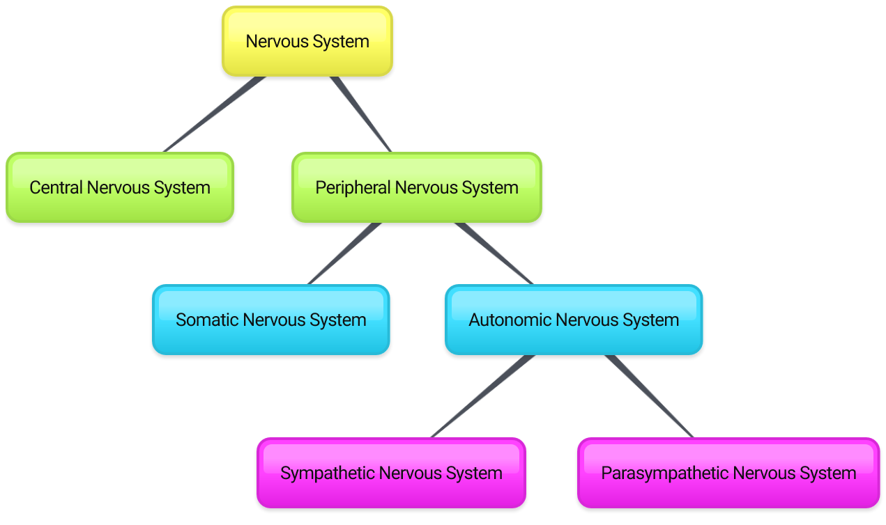
The PNS is divided into two major parts, called the autonomic and somatic nervous systems. The somatic nervous system (SNS) is responsible for activities that are under voluntary control and awareness, such as turning a steering wheel or feeling water splashing on your hands. The autonomic nervous system (ANS) controls involuntary activities, such as digesting a meal or regulating your heartbeat. The autonomic nervous system also has two divisions: the sympathetic and parasympathetic nervous systems. The sympathetic nervous system controls the "fight-or-flight" response during emergencies, and the parasympathetic nervous system controls the routine “housekeeping” functions of the body (sometimes referred to as the "rest and digest" division). Generally speaking, your body is usually activating a balance of sympathetic and parasympathetic functions.
The CNS and the sympathetic and parasympathetic branches of the ANS (itself a division of the PNS) are described in greater detail in other modules of this chapter. The other division of the PNS, the SNS, is composed largely of sensory receptors and their connections (covered in the sensory system chapter) and structures related to voluntary motor control (addressed in the movement chapter).
Central Nervous System Regions
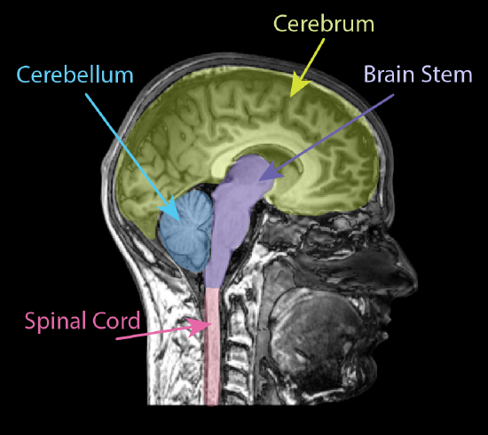
As mentioned previously, the CNS is composed of the brain and spinal cord. In its fully formed state, the CNS may be further broken down into four major regions: the cerebrum, the brainstem, the cerebellum, and the spinal cord. The folded outer tissue of the cerebrum is what most people envision when they think of "the brain." Although this is a major component, there are several other critical structures that make up the rest of the brain including those in the brainstem and cerebellum. The brainstem itself is comprised of several structures from all of the main subdivisions of the brain. All of these major CNS regions are colored in on an MRI of an adult in Figure \(\PageIndex{2}\).
The brain is often considered as the most interesting and diversified part of the CNS. Hundreds of specific brain regions with distinct structures and functions have been identified over many years of scientific study. Many of the commonly used terms for many regions of the brain, such as forebrain, midbrain, and hindbrain derive from embryonic development. The origins of these terms and details of brain development are in the supplemental section on the embryonic development of the CNS which appears later in this text.
Furthermore, the overall organization, specific structural characteristics, and the main functions of several key regions of the brain and spinal cord will be detailed in the next module of this chapter. For now, see Figure \(\PageIndex{3}\) below to provide a framework for how the various organizational schemes overlap. For most, separating the brain into the Forebrain, Midbrain, and Hindbrain is the easiest to remember.

Protective Coverings of the Central Nervous System
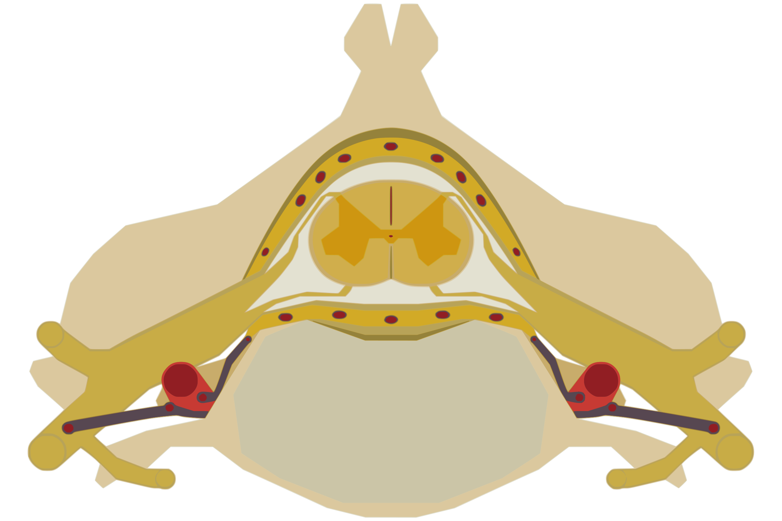
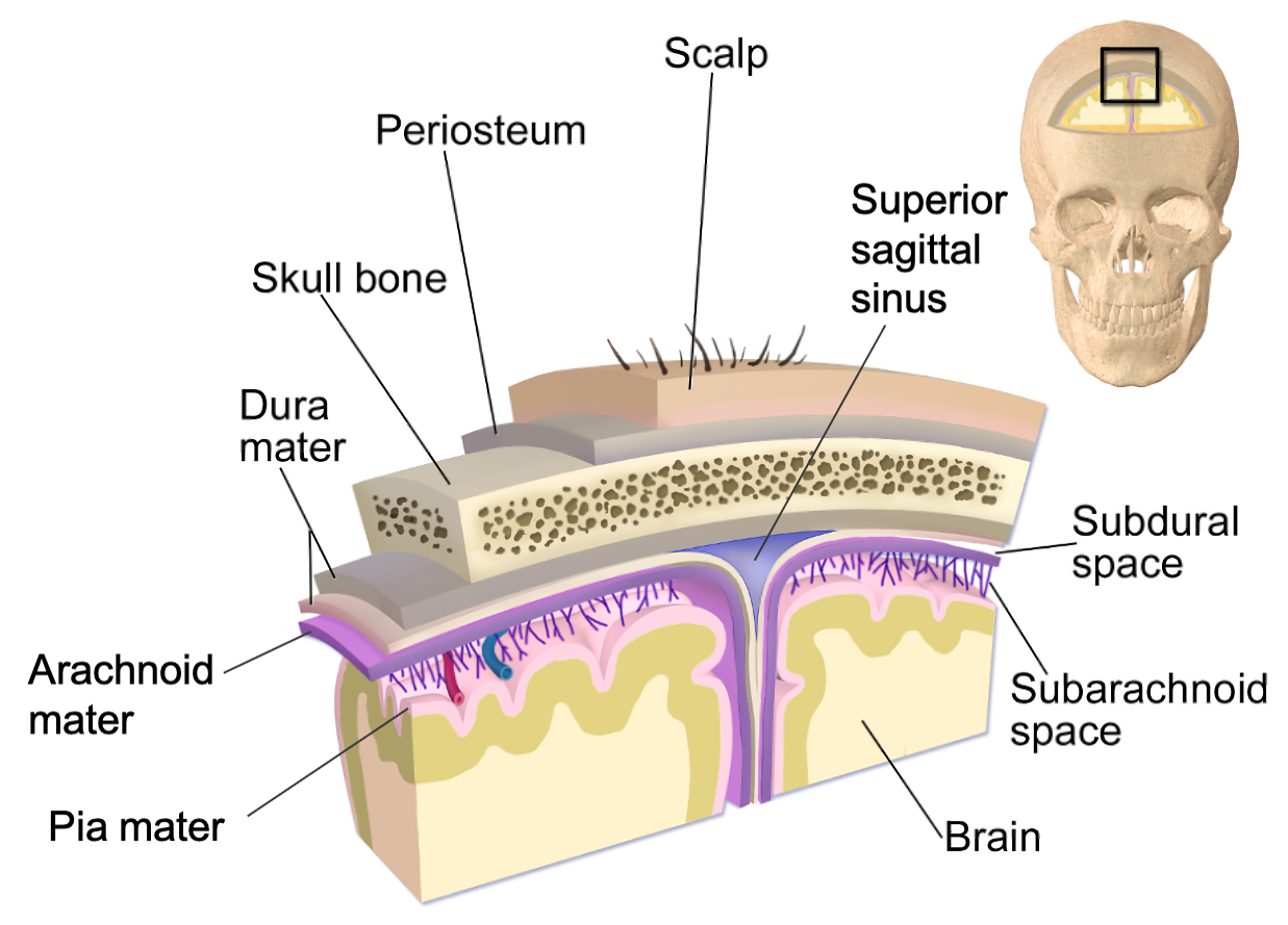
The central nervous system (CNS) is crucial to the operation of the body and any compromise of function in the brain and spinal cord can lead to severe difficulties. Both the brain and spinal cord are protected by multiple structures. First, the bones of the skull enclose and house the brain, and the bones of the vertebral column enclose and protect the spinal cord. Figure \(\PageIndex{4}\) shows a segment of the spinal cord within its protective vertebra.
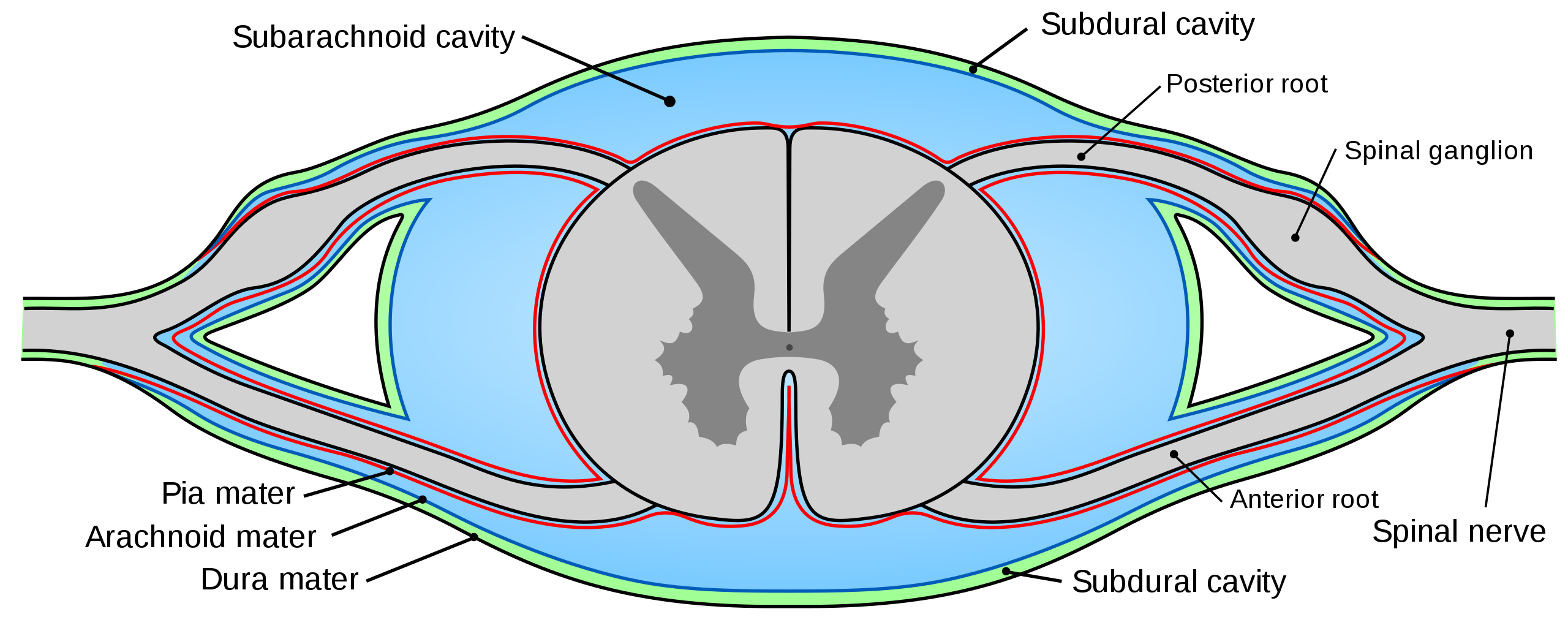
Underneath the skeletal structures, the brain and spinal cord are surrounded and protected by tough meninges, a three-layer protective sheath made of connective tissue that surrounds, supports, stabilizes and partitions the nervous tissue. The meninges also contain cushioning cerebrospinal fluid (CSF). The three meningeal layers are the dura mater, the arachnoid mater, and the pia mater. Figure \(\PageIndex{5}\) shows the three layers of meninges covering the spinal cord and Figure \(\PageIndex{6}\) shows all the protective structures of the brain. The subdural cavity of the spinal cord and the subdural space of the brain lie between the dura mater and the arachnoid mater, whereas the subarachnoid cavity of the spinal cord and the subarachnoid space of the brain lie between the arachnoid mater and the pia mater. The superior sagittal sinus is a space between layers of dura mater immediately superior to the longitudinal fissure (the separation between the left and right hemispheres) of the brain.
In the spinal cord, the posterior (/dorsal) and anterior (/ventral) roots converge to form the spinal nerve. The spinal ganglion (also known as the dorsal root ganglion), an enlargement of the posterior (/dorsal) root, encases sensory neuron cell bodies.
Ventricular System
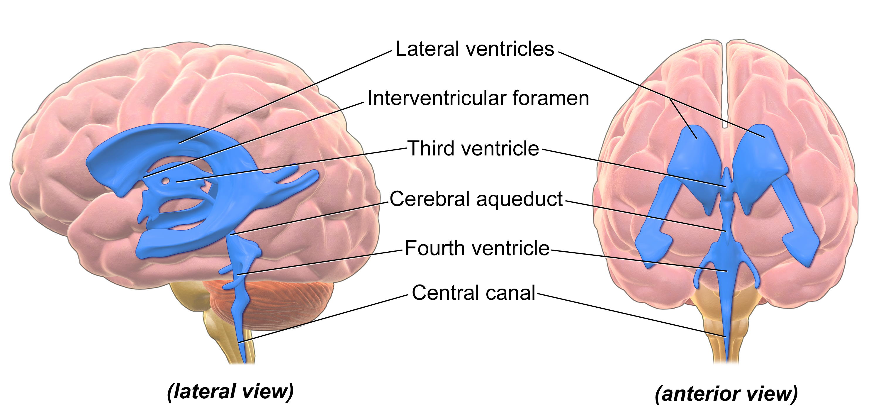
There are four ventricles within the brain, all of which developed from the original hollow space within the neural tube, the central canal. The ventricles are lined with ependymal cells (a type of glia) that produce cerebrospinal fluid (CSF). The first two ventricles are named the lateral ventricles and are deep within the brain. The two lateral ventricles are shaped like a letter "C" and are located in the left and right hemispheres. (They were originally referred to as the first and second ventricles.) These ventricles are connected to the third ventricle by two openings called the interventricular foramina (plural; "foramen" is the singular term). The third ventricle opens into a canal called the cerebral aqueduct that passes through the midbrain and connects the third ventricle to the fourth ventricle. The fourth ventricle is the space between the cerebellum and the pons and upper medulla. From the fourth ventricle, CSF continues down the central canal of the spinal cord. Figure \(\PageIndex{7}\) illustrates the ventricles, from both a lateral and anterior viewpoint. CSF circulates through the brain and the spinal cord within these structures.
Summary
The organization of the nervous system includes six divisions. The CNS includes the brain and spinal cord, and the PNS consists of all other nervous tissue in the body. The nerves of the PNS connect the CNS to the rest of the body. The PNS is further divided into the autonomic and somatic nervous systems. The somatic nervous system (SNS) is responsible for activities that are under voluntary control and awareness. The autonomic nervous system (ANS) controls involuntary activities. The autonomic nervous system also has two divisions: the sympathetic and parasympathetic nervous systems. The sympathetic nervous system controls response during emergencies, and the parasympathetic nervous system controls the routine functions of the body.
The adult CNS is separated into four major regions: the cerebrum, the brainstem, the cerebellum, and the spinal cord. Anatomical structures help to protect and isolate the CNS, including the skull (surrounding the brain) and the vertebral column (surrounding the spinal cord). Layers of connective tissue called meninges support and stabilize the brain and spinal cord, as well as partition the brain into specific regions. The outer layer is the dura mater, the middle layer is the arachnoid mater and the inner layer is the pia mater.
The ventricular system is composed of four fluid-filled ventricles in the brain (two lateral ventricles, the third ventricle and the fourth ventricle) which connect to each other (via the interventricular foramina and the cerebral aqueduct) and the central canal of the spinal cord. Cerebrospinal fluid (CSF) circulates through the brain and the spinal cord within these structures.
Additional Resources
If you are curious about anatomical terms used to describe different areas of the body, see "Anatomical Vocabulary" on: " Anatomical Position and Planes" by LibreTexts, licensed under CC BY-SA .
For more detail on the embryological development of the brain and the primary and secondary brain vesicles, see: " The Embryologic Perspective" by OpenStax, LibreTexts, licensed under CC BY .
For more detail on the meninges and the ventricles, see: " Support and Protection of the Brain" by Whitney Menefee, Julie Jenks, Chiara Mazzasette, & Kim-Leiloni Nguyen, LibreTexts, licensed under CC BY and " Circulation and the Central Nervous System" by OpenStax, LibreTexts, licensed under CC BY .
Attributions
- Figures:
- Nervous System Flowchart by Suzanne Wakim dedicated CC0. From " Introduction to the Nervous System" by Suzanne Wakim & Mandeep Grewal, LibreTexts is licensed under CC BY-NC .
- Adapted by Suzanne Wakim from MRI brain sagittal section.jpg by everyone's idle, licensed CC BY-SA 2.0 via Wikimedia Commons.
- Time3000 is licensed under CC BY-SA 2.5 via Wikimedia Commons
- Mysid, Public domain, via Wikimedia Commons
- "Cranial meninges" by Chiara Mazzasette is licensed under CC BY 4.0 / A derivative of Blausen 0110 BrainLayers.png
- "Blausen 0896 Ventricles Brain" by BruceBlaus is in the Public Domain, CC0
- Text adapted from:
- " Anatomical Position and Planes" by LibreTexts is licensed under CC BY-SA .
- " Introduction to the Nervous System" by Suzanne Wakim & Mandeep Grewal, LibreTexts is licensed under CC BY-NC .
- " The Central Nervous System" by OpenStax, LibreTexts is licensed under CC BY .
- " Support and Protection of the Brain" by Whitney Menefee, Julie Jenks, Chiara Mazzasette, & Kim-Leiloni Nguyen, LibreTexts is licensed under CC BY .
- " Central Nervous System" by Suzanne Wakim & Mandeep Grewal, LibreTexts is licensed under CC BY-NC .
- Changes: Text (and most of the images) from above four sources pieced together with some modifications, transitions and additional content (and images) added by Naomi I. Gribneau Bahm, PhD., Psychology Professor at Cosumnes River College, Sacramento, CA. and Alan S. Keys, Ph.D, Psychology Professor at Sacramento City College, Sacramento, CA.
- Additional revisions made by Alan Keys, Ph.D., Sacramento City College, Sacramento, CA.