Social Sci LibreTexts

3: Molecular Biology and Genetics

  • Page ID
    66707

    \( \newcommand{\vecs}[1]{\overset { \scriptstyle \rightharpoonup} {\mathbf{#1}} } \)

    \( \newcommand{\vecd}[1]{\overset{-\!-\!\rightharpoonup}{\vphantom{a}\smash {#1}}} \)

    \( \newcommand{\id}{\mathrm{id}}\) \( \newcommand{\Span}{\mathrm{span}}\)

    ( \newcommand{\kernel}{\mathrm{null}\,}\) \( \newcommand{\range}{\mathrm{range}\,}\)

    \( \newcommand{\RealPart}{\mathrm{Re}}\) \( \newcommand{\ImaginaryPart}{\mathrm{Im}}\)

    \( \newcommand{\Argument}{\mathrm{Arg}}\) \( \newcommand{\norm}[1]{\| #1 \|}\)

    \( \newcommand{\inner}[2]{\langle #1, #2 \rangle}\)

    \( \newcommand{\Span}{\mathrm{span}}\)

    \( \newcommand{\id}{\mathrm{id}}\)

    \( \newcommand{\Span}{\mathrm{span}}\)

    \( \newcommand{\kernel}{\mathrm{null}\,}\)

    \( \newcommand{\range}{\mathrm{range}\,}\)

    \( \newcommand{\RealPart}{\mathrm{Re}}\)

    \( \newcommand{\ImaginaryPart}{\mathrm{Im}}\)

    \( \newcommand{\Argument}{\mathrm{Arg}}\)

    \( \newcommand{\norm}[1]{\| #1 \|}\)

    \( \newcommand{\inner}[2]{\langle #1, #2 \rangle}\)

    \( \newcommand{\Span}{\mathrm{span}}\) \( \newcommand{\AA}{\unicode[.8,0]{x212B}}\)

    \( \newcommand{\vectorA}[1]{\vec{#1}}      % arrow\)

    \( \newcommand{\vectorAt}[1]{\vec{\text{#1}}}      % arrow\)

    \( \newcommand{\vectorB}[1]{\overset { \scriptstyle \rightharpoonup} {\mathbf{#1}} } \)

    \( \newcommand{\vectorC}[1]{\textbf{#1}} \)

    \( \newcommand{\vectorD}[1]{\overrightarrow{#1}} \)

    \( \newcommand{\vectorDt}[1]{\overrightarrow{\text{#1}}} \)

    \( \newcommand{\vectE}[1]{\overset{-\!-\!\rightharpoonup}{\vphantom{a}\smash{\mathbf {#1}}}} \)

    \( \newcommand{\vecs}[1]{\overset { \scriptstyle \rightharpoonup} {\mathbf{#1}} } \)

    \( \newcommand{\vecd}[1]{\overset{-\!-\!\rightharpoonup}{\vphantom{a}\smash {#1}}} \)

    \(\newcommand{\avec}{\mathbf a}\) \(\newcommand{\bvec}{\mathbf b}\) \(\newcommand{\cvec}{\mathbf c}\) \(\newcommand{\dvec}{\mathbf d}\) \(\newcommand{\dtil}{\widetilde{\mathbf d}}\) \(\newcommand{\evec}{\mathbf e}\) \(\newcommand{\fvec}{\mathbf f}\) \(\newcommand{\nvec}{\mathbf n}\) \(\newcommand{\pvec}{\mathbf p}\) \(\newcommand{\qvec}{\mathbf q}\) \(\newcommand{\svec}{\mathbf s}\) \(\newcommand{\tvec}{\mathbf t}\) \(\newcommand{\uvec}{\mathbf u}\) \(\newcommand{\vvec}{\mathbf v}\) \(\newcommand{\wvec}{\mathbf w}\) \(\newcommand{\xvec}{\mathbf x}\) \(\newcommand{\yvec}{\mathbf y}\) \(\newcommand{\zvec}{\mathbf z}\) \(\newcommand{\rvec}{\mathbf r}\) \(\newcommand{\mvec}{\mathbf m}\) \(\newcommand{\zerovec}{\mathbf 0}\) \(\newcommand{\onevec}{\mathbf 1}\) \(\newcommand{\real}{\mathbb R}\) \(\newcommand{\twovec}[2]{\left[\begin{array}{r}#1 \\ #2 \end{array}\right]}\) \(\newcommand{\ctwovec}[2]{\left[\begin{array}{c}#1 \\ #2 \end{array}\right]}\) \(\newcommand{\threevec}[3]{\left[\begin{array}{r}#1 \\ #2 \\ #3 \end{array}\right]}\) \(\newcommand{\cthreevec}[3]{\left[\begin{array}{c}#1 \\ #2 \\ #3 \end{array}\right]}\) \(\newcommand{\fourvec}[4]{\left[\begin{array}{r}#1 \\ #2 \\ #3 \\ #4 \end{array}\right]}\) \(\newcommand{\cfourvec}[4]{\left[\begin{array}{c}#1 \\ #2 \\ #3 \\ #4 \end{array}\right]}\) \(\newcommand{\fivevec}[5]{\left[\begin{array}{r}#1 \\ #2 \\ #3 \\ #4 \\ #5 \\ \end{array}\right]}\) \(\newcommand{\cfivevec}[5]{\left[\begin{array}{c}#1 \\ #2 \\ #3 \\ #4 \\ #5 \\ \end{array}\right]}\) \(\newcommand{\mattwo}[4]{\left[\begin{array}{rr}#1 \amp #2 \\ #3 \amp #4 \\ \end{array}\right]}\) \(\newcommand{\laspan}[1]{\text{Span}\{#1\}}\) \(\newcommand{\bcal}{\cal B}\) \(\newcommand{\ccal}{\cal C}\) \(\newcommand{\scal}{\cal S}\) \(\newcommand{\wcal}{\cal W}\) \(\newcommand{\ecal}{\cal E}\) \(\newcommand{\coords}[2]{\left\{#1\right\}_{#2}}\) \(\newcommand{\gray}[1]{\color{gray}{#1}}\) \(\newcommand{\lgray}[1]{\color{lightgray}{#1}}\) \(\newcommand{\rank}{\operatorname{rank}}\) \(\newcommand{\row}{\text{Row}}\) \(\newcommand{\col}{\text{Col}}\) \(\renewcommand{\row}{\text{Row}}\) \(\newcommand{\nul}{\text{Nul}}\) \(\newcommand{\var}{\text{Var}}\) \(\newcommand{\corr}{\text{corr}}\) \(\newcommand{\len}[1]{\left|#1\right|}\) \(\newcommand{\bbar}{\overline{\bvec}}\) \(\newcommand{\bhat}{\widehat{\bvec}}\) \(\newcommand{\bperp}{\bvec^\perp}\) \(\newcommand{\xhat}{\widehat{\xvec}}\) \(\newcommand{\vhat}{\widehat{\vvec}}\) \(\newcommand{\uhat}{\widehat{\uvec}}\) \(\newcommand{\what}{\widehat{\wvec}}\) \(\newcommand{\Sighat}{\widehat{\Sigma}}\) \(\newcommand{\lt}{<}\) \(\newcommand{\gt}{>}\) \(\newcommand{\amp}{&}\) \(\definecolor{fillinmathshade}{gray}{0.9}\)

    Hayley Mann, M.A., Binghamton University

    Xazmin Lowman, Ph.D., University of California, Irvine

    Malaina Gaddis, Ph.D.

    Learning Objectives

    • Define terms useful to molecular biology and genetics.
    • Explain and identify the purpose of both DNA replication and the cell cycle.
    • Identify key differences between mitosis and meiosis.
    • Outline the process of protein synthesis including transcription and translation.
    • Use principles of Mendelian inheritance to predict genotypes and phenotypes of future generations.
    • Explain complexities surrounding patterns of genetic inheritance and polygenic traits.
    • Discuss challenges to and bioethical concerns of genetic testing.

    I [Hayley Mann] started my Bachelor’s degree in 2003, which was the same year the Human Genome Project released its first draft sequence. I initially declared a genetics major because I thought it sounded cool. However, upon taking an actual class, I discovered that genetics was challenging. In addition to my genetics major, I signed up for biological anthropology classes and soon learned that anthropology could bring all those molecular lessons to life. For instance, we are composed of cells, proteins, nucleic acids, carbohydrates, and lipids. Anthropologists often include these molecules in their studies to identify how humans vary; if there are meaningful differences, they propose theories to explain them.

    Since the release of the first human genome sequence, the field of genetics has grown into genomics. Researchers now address these complex questions on a large scale. To process “big data,” some scientists have moved to working on a computer full time doing computational biology. As you learned in Chapter 1, molecular anthropologists use genetics to compare ancient and modern populations as well as study nonhuman primates. Molecular anthropologists must also stay current with advancing technology (you will learn about the results of some of this genomic research as it has been applied to fossils in Chapters 11 and 12). If you wish to be part of this dynamic field, then take advantage of available campus laboratory classes and internships and also never stop reading scientific papers.

    This chapter provides the basics for understanding human variation and how the evolutionary process works. A few advanced genetics topics are also presented because biotechnology is now commonplace in health and society. Understanding the science behind this remarkable field means you will be able to participate in bioethical and anthropological discussions as well as make more informed decisions regarding genetic testing.

    About the Authors

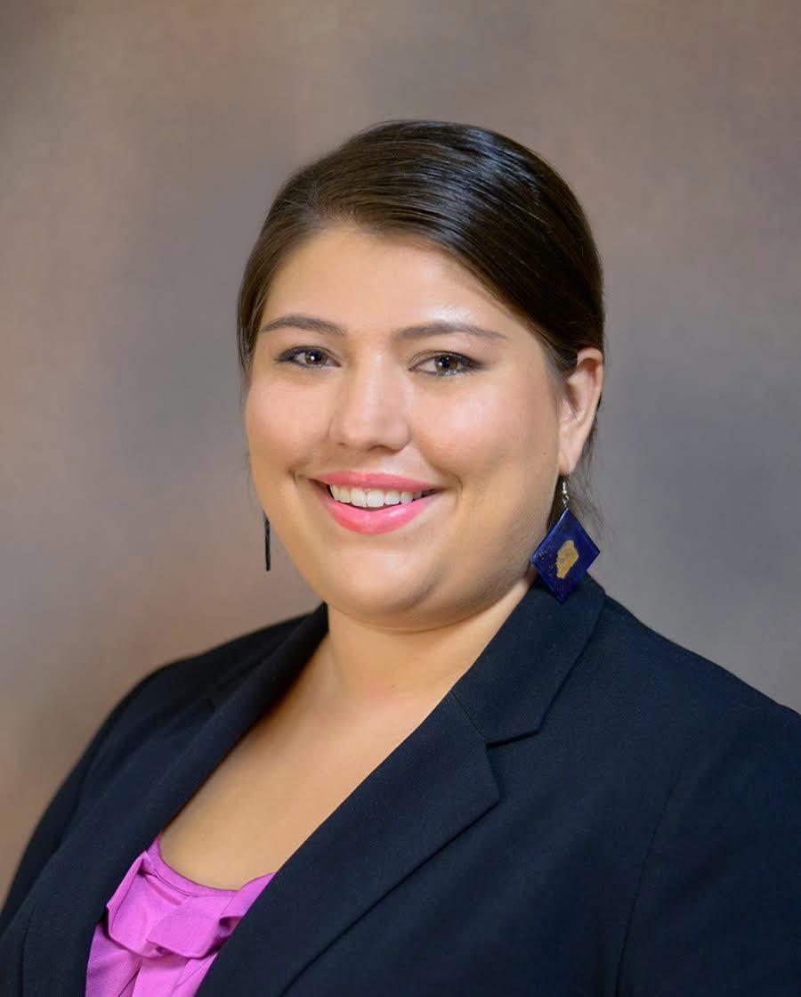
    Hayley Mann, M.A.

    Binghamton University, hmann3@binghamton.edu

    alt
    Hayley Mann

    Hayley Mann received her bachelor’s degree in Genetics from the University of California, Davis, and continued her graduate studies in Biological and Molecular Anthropology at the California State University, Sacramento. She is currently a Ph.D. candidate at Binghamton University, where her dissertation focus is on studying genetic variation of Pacific Islanders (Republic of Vanuatu) and also changes in health as the result of colonization. Hayley also works in clinical genomics and specializes in various DNA-sequencing methodologies.

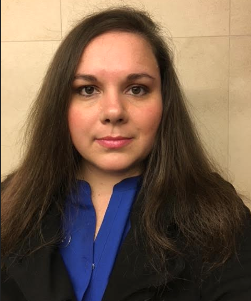
    Xazmin Lowman, Ph.D.

    University of California, Irvine, xlowman@uci.edu

    alt
    Xazmin Lowman

    Xazmin Lowman received her bachelor’s degree in Physiology from the University of Arizona. She pursued her doctorate in cancer biology at the University of Minnesota, where she studied how blood cancers evade cell death. Currently, her scientific interests have led to a postdoctoral fellowship at the University of California, Irvine, where she studies how solid tumors adapt to metabolic stress. Beyond figuring out the mechanisms that drive cancer, she enjoys traveling to learn of other cultures and share her own upbringing on the Navajo reservation.

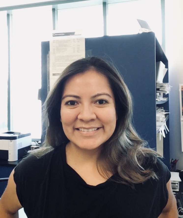
    Malaina Gaddis, Ph.D.

    malainagaddis@gmail.com

    alt
    Malaina Gaddis

    Malaina Gaddis studied biology at Brigham Young University in Utah and then earned a Ph.D. in genetics and molecular and cellular biology at the University of Southern California. During her Ph.D. studies, she investigated small molecule inhibitors that alter epigenetic modifications and gene expression in cancer cells. Following her Ph.D., she focused on the business side of the biotech industry by completing a master’s degree in bioscience management at the Keck Graduate Institute. Malaina is especially interested in using genomics to further genetic testing and personalized treatment and currently works as a genome variation scientist.

    For Further Exploration

    Websites

    National Human Genome Research Institute https://www.genome.gov/

    Genetics Home Reference https://ghr.nlm.nih.gov/

    Genetics Generation http://knowgenetics.org/

    yourgenome https://www.yourgenome.org/

    Cardiovascular Disease: Genes are Important, but Health-Related Behaviors and Lifestyle Choices Can Make or Break Your Health http://ehrweb.aaas.org/ehr/books/3_howard.html

    Gene Sequencing Speeds Diagnosis of Deadly Newborn Diseases http://www.pbs.org/wgbh/nova/next/body/newborn-gene-sequencing/

    Carl Zimmer’s Game of Genomes https://www.statnews.com/feature/game-of-genomes/season-one/

    Illumina Sequencing by Synthesis https://www.youtube.com/watch?v=fCd6B5HRaZ8

    Articles

    Aartsma-Rus, Annemieke, Ieke B. Ginjaar, and Kate Bushby. 2016. “The Importance of Genetic Diagnosis for Duchenne Muscular Dystrophy.” Journal of Medical Genetics 53 (3): 145–151.

    Acuna-Hidalgo, Rocio, Joris A. Veltman, and Alexander Hoischen. 2016. “New Insights into the Generation and Role of De Novo Mutations in Health and Disease.” Genome Biology 17 (241): 1-19.

    Albert, Benjamin, Susanna Tomassetti, Yvonne Gloor, Daniel Dilg, Stefano Mattarocci, Slawomir Kubik, Lukas Hafner, and David Shore. 2019. “Sfp1 Regulates Transcriptional Networks Driving Cell Growth and Division through Multiple Promoter-Binding Modes.” Genes & Development 33 (5–6): 288–293.

    Almathen, Faisal, Haitham Elbir, Hussain Bahbahani, Joram Mwacharo, and Olivier Hanotte. 2018. “Polymorphisms in Mc1r and Asip Genes Are Associated With Coat Color Variation in the Arabian Camel.” Journal of Heredity 109 (6): 700–706.

    Ballester, Leomar Y., Rajyalakshmi Luthra, Rashmi Kanagal-Shamanna, and Rajesh R. Singh. 2016. “Advances in Clinical Next-Generation Sequencing: Target Enrichment and Sequencing Technologies.” Expert Review of Molecular Diagnostics 16 (3): 357–372.

    Baranovskiy, Andrey G., Vincent N. Duong, Nigar D. Babayeva, Yinbo Zhang, Youri I. Pavlov, Karen S. Anderson, and Tahir H. Tahirov. 2018. “Activity and Fidelity of Human DNA Polymerase Alpha Depend on Primer Structure.” Journal of Biological Chemistry 293 (18): 6,824–6,843.

    Brezina, Paulina R., Raymond Anchan, and William G. Kearns. 2016. “Preimplantation Genetic Testing for Aneuploidy: What Technology Should You Use and What Are the Differences?” Journal of Assisted Reproduction and Genetics 33 (7): 823–832.

    Bultman, Scott J. 2017. “Interplay Between Diet, Gut Microbiota, Epigenetic Events, and Colorectal Cancer.” Molecular Nutrition & Food Research 61 (1):1-12.

    Cutting, Garry R. 2015. “Cystic Fibrosis Genetics: From Molecular Understanding to Clinical Application.” Nature Reviews Genetics 16 (1): 45–56.

    D’Alessandro, Giuseppina., and Fabrizio d’Adda di Fagagna. 2017. “Transcription and DNA Damage: Holding Hands or Crossing Swords?” Journal of Molecular Biology 429 (21): 3,215–3,229.

    De Craene, Johan-Owen, Dimitri L. Bertazzi, Séverine Bar, and Sylvie Friant. 2017. “Phosphoinositides, Major Actors in Membrane Trafficking and Lipid Signaling Pathways.” International Journal of Molecular Sciences 18 (3): 1-20.

    Deng, Lian, and Shuhua Xu. 2018. “Adaptation of Human Skin Color in Various Populations.” Hereditas 155 (1): 1-12.

    Dever, Thomas E., Terri G. Kinzy, and Graham D. Pavitt. 2016. “Mechanism and Regulation of Protein Synthesis in Saccharomyces Cerevisiae.” Genetics 203 (1): 65–107.

    Eme, Laura, Anja Spang, Jonathan Lombard, Courtney W. Stairs, and Thijs J. G. Ettema. 2017. “Archaea and the Origin of Eukaryotes.” Nature Reviews Microbiology 15 (12): 711–723.

    Gomez-Carballa, Alberto, Jacobo Pardo-Seco, Stefania Brandini, Alessandro Achilli, Ugo A. Perego, Michael D. Coble, Toni M. Diegoli, et al. 2018. “The Peopling of South America and the Trans-Andean Gene Flow of the First Settlers.” Genome Research 28 (6): 767–779.

    Gvozdenov, Zlata, Janhavi Kolhe, and Brian C. Freeman. 2019. “The Nuclear and DNA-Associated Molecular Chaperone Network.” Cold Spring Harbor Perspectives in Biology. New York:Cold Spring Harbor Laboratory Press.

    Harkins, Kelly M., and Anne C. Stone. 2015. “Ancient Pathogen Genomics: Insights Into Timing and Adaptation.” Journal of Human Evolution 79: 137–149.

    Jackson, Maria, Leah Marks, Gerhard H. W. May, and Joanna B. Wilson. 2018. “The Genetic Basis of Disease.” Essays in Biochemistry 62 (5): 643–723.

    Lenormand, Thomas., Jan Engelstadter, Susan E. Johnston, Erik Wijnker, and Christopher R. Haag. 2016. “Evolutionary Mysteries in Meiosis.” Philosophical Transactions of the Royal Society B 371: 1-14.

    Levy, Shawn E., and Richard M. Myers. 2016. “Advancements in Next-Generation Sequencing.” Annual Review of Genomics and Human Genetics 17: 95–115.

    Lu, Mengfei, Cathryn M. Lewis, and Matthew Traylor. 2017. “Pharmacogenetic Testing Through the Direct-to-Consumer Genetic Testing Company 23andme.” BMC Medical Genomics 10 (47): 1-8.

    Ly, Lundi, Donovan Chan, Mahmoud Aarabi, Mylene Landry, Nathalie A. Behan, Amanda J. MacFarlane, and Jacquetta Trasler. 2017. “Intergenerational Impact of Paternal Lifetime Exposures to Both Folic Acid Deficiency and Supplementation on Reproductive Outcomes and Imprinted Gene Methylation.” Molecular Human Reproduction 23 (7): 461–477.

    Ma, Wenxiu, Giancarlo Bonora, Joel B. Berletch, Xinxian Deng, William S. Noble, and Christine M. Disteche. 2018. “X-Chromosome Inactivation and Escape From X Inactivation in Mouse.” Methods in Molecular Biology 1,861: 205–219.

    Machiela, Mitchell J., Weiyin Zhou, Eric Karlins, Joshua N. Sampson, Neal D. Freedman, Qi Yang, Belynda Hicks, et al. 2016. “Female Chromosome X Mosaicism Is Age-Related and Preferentially Affects the Inactivated X Chromosome.” Nat Commun 7: 1-9. doi: 10.1038/ncomms11843.

    Mahdavi, Morteza, Mohammadreza Nassiri, Mohammad M. Kooshyar, Masoume Vakili-Azghandi, Amir Avan, Ryan Sandry, Suja Pillai, Alfred K. Lam, and Vinod Gopalan. 2019. “Hereditary Breast Cancer; Genetic Penetrance and Current Status With BRCA.” Journal of Cellular Physiology 234 (5): 5,741–5,750.

    McDade, Thomas W., Calen P. Ryan, Meaghan J. Jones, Morgan K. Hoke, Judith Borja, Gregory E. Miller, Christopher W. Kuzawa, and Michael S. Kobor. 2019. “Genome-Wide Analysis of DNA Methylation in Relation to Socioeconomic Status During Development and Early Adulthood.” American Journal of Physical Anthropology 169 (1): 3–11.

    Migeon, Barbara R. 2017. “Choosing the Active X: The Human Version of X Inactivation.” Trends in Genetics 33 (12): 899–909.

    Myerowitz, Rachel. 1997. “Tay-Sachs Disease-Causing Mutations and Neutral Polymorphisms in the Hex a Gene.” Human Mutation 9: 195–208.

    Onufriev, Alexey V. and Helmut Schiessel. 2019. “The Nucleosome: From Structure to Function Through Physics.” Current Opinion in Structural Biology 56: 119–130.

    Quillen, Ellen E., Heather L. Norton, Esteban J. Parra, Frida Lona-Durazo, Khai C. Ang, Florin M. Illiescu, Laurel N. Pearson, et al. 2019. “Shades of Complexity: New Perspectives on the Evolution and Genetic Architecture of Human Skin.” American Journal of Physical Anthropology 168 (67): 4–26.

    Raspelli, Erica and Roberta Fraschini. 2019. “Spindle Pole Power in Health and Disease.” Current Genetics 65 (4): 851-855.

    Ravinet, M., R. Faria, R. K. Butlin, J. Galindo, N. Bierne, M. Rafajlovic, M. A. F. Noor, B. Mehlig, and A. M. Westram. 2017. “Interpreting the Genomic Landscape of Speciation: A Road Map for Finding Barriers to Gene Flow.” Journal of Evolutionary Biology 30 (8): 1,450–1,477.

    Regev, Aviv, Sarah A. Teichmann, Eric S. Lander, Ido Amit, Christophe Benoist, Ewan Birney, Bernd Bodenmiller, et al. 2017. “The Human Cell Atlas.” Elife 6e27041: 1-30. doi: doi.org/10.7554.eLife.27041.

    Roberts, Andrea L., Nicole Gladish, Evan Gatev, Meaghan J. Jones, Ying Chen, Julia L. MacIsaac, Shelley S. Tworoger, et al. 2018. “Exposure to Childhood Abuse Is Associated With Human Sperm DNA Methylation.” Translational Psychiatry 8 (194): 1-11.

    Roger, Andrew J., Sergio A. Muñoz-Gómez, and Ryoma Kamikawa. 2017. “The Origin and Diversification of Mitochondria.” Current Biology 27 (21): R1177–R1192.

    Ségurel, Laure and Céline Bon. 2017. “On the Evolution of Lactase Persistence in Humans.” Annual Review of Genomics and Human Genetics 18: 297–319.

    Sheth, Bhavisha P. and Vrinda S. Thaker. 2017. “DNA Barcoding and Traditional Taxonomy: An Integrated Approach for Biodiversity Conservation.” Genome 60 (7): 618–628.

    Snedeker, Jonathan, Matthew Wooten, and Xin Chen. 2017. “The Inherent Asymmetry of DNA Replication.” Annual Review of Cell and Developmental Biology 33: 291–318.

    Sullivan-Pyke, Chantae and Anuja Dokras. 2018. “Preimplantation Genetic Screening and Preimplantation Genetic Diagnosis.” Obstetrics and Gynecology Clinics of North America 45 (1): 113–125.

    Szostak, Jack W. 2017. “The Narrow Road to the Deep Past: In Search of the Chemistry of the Origin of Life.” Angewandte Chemie International Edition 56 (37): 11,037–11,043.

    Tessema, Mathewos, Ulrich Lehmann, and Hans Kreipe. 2004. “Cell Cycle and No End.” Virchows Archiv European Journal of Pathology 444 (4): 313–323.

    Tishkoff, Sarah A., Floyd A. Reed, Alessia Ranciaro, Benjamin F. Voight, Courtney C. Babbitt, Jesse S. Silverman, Kweli Powell, et al. 2007. “Convergent Adaptation of Human Lactase Persistence in Africa and Europe.” Nature Genetics 39 (1): 31–40.

    Visootsak, Jeannie and John M. Graham, Jr. 2006. “Klinefelter Syndrome and Other Sex Chromosomal Aneuploidies.” Orphanet Journal of Rare Diseases 1:42 doi: 10.1186/1750-1172-1-42.

    Yamamoto, Fumi-ichiro, Henrik Clausen, Thayer White, John Marken, and Sen-itiroh Hakomori. 1990. “Molecular Genetic Basis of the Histo-Blood Group Abo System.” Nature 345: 229–233.

    Zlotogora, Joël. 2003. “Penetrance and Expressivity in the Molecular Age.” Genetics in Medicine 5: 347–352.

    Zorina-Lichtenwalter, Katerina, Ryan N. Lichtenwalter, Dima V. Zaykin, Marc Parisien, Simon Gravel, Andrey Bortsov, and Luda Diatchenko. 2019. “A Study in Scarlet: MC1R as the Main Predictor of Red Hair and Exemplar of the Flip-Flop Effect.” Human Molecular Genetics 28 (12): 2,093-2,106.

    Zwart, Haeh. 2018. “In the Beginning Was the Genome: Genomics and the Bi-Textuality of Human Existence.” New Bioethics 24 (1): 26–43.
    References

    Lindo, John, Emilia Huerta-Sánchez, Shigeki Nakagome, Morten Rasmussen, Barbara Petzelt, Joycelynn Mitchell, Jerome S. Cybulski,et al. 2016. “A Time Transect of Exomes From a Native American Population Before and After European Contact.” Nature Communications 7: 1–11. doi: 10.1038/ncomms13175.

    Skloot, Rebecca. 2010. The Immortal Life of Henrietta Lacks. New York: Crown Publishing Group.

    Wolfe, George C., dir. 2017. The Immortal Life of Henrietta Lacks. HBO Films, April 22, 2017. TV Movie.

    Figure Attributions

    Figure 3.1 Phospholipid Bilayer (Anatomy & Physiology, Chapter 3.1, Figure 2) by OpenStax is used under a CC BY 4.0 License.

    Figure 3.2 Nucleic Acid by NIH National Human Genome Research Institute is in the public domain.

    Figure 3.3 Amino acids (Biology 2e, Figure 3.22) by OpenStax is used under a CC BY 4.0 License.

    Figure 3.4 Amino Acids by NIH National Human Genome Research Institute is in the public domain.

    Figure 3.5 Bacteria by NIH National Human Genome Research Institute is in the public domain.

    Figure 3.6 Bryum capillare lamina by Kristian Peters [Fabelfroh 13:07, 2007 (UTC)] is used under a CC BY-SA 3.0 License.

    Figure 3.7 Cell Membrane (Anatomy & Physiology, Figure 3.4) by OpenStax is used under a CC BY 4.0 License.

    Figure 3.8 Organelle by NIH National Human Genome Research Institute is in the public domain.

    Figure 3.9 Nucleus (Biology 2e, Figure 4.11) by OpenStax is used under a CC BY 4.0 License.

    Figure 3.10 Mitochondrion through an electron microscope (Biology 2e, Figure 4.14) by OpenStax is used under a CC BY 4.0 License.

    Figure 3.11 Cell Structure table original to Explorations: An Open Invitation to Biological Anthropology by Hayley Mann, Xazmin Lowman, and Malaina Gaddis is under a CC BY-NC 4.0 License.

    Figure 3.12a A group of Tsimshian people having a tea party in a tent, Lax Kw’alaams (formerly Port Simpson), B.C., c. 1890 by unknown photographer at Online MIKAN no. 3368729 Library and Archives Canada (C-060817). Copyright is expired.

    Figure 3.12b Tsimshian Territory map original to Explorations: An Open Invitation to Biological Anthropology by Elyssa Ebding at GeoPlace, California State University, Chico is under a CC BY-NC 4.0 License.

    Figure 3.13 Rosalind Franklin from the personal collection of Jenifer Glynn by MRC Laboratory of Molecular Biology is used under a CC BY-SA 4.0 License.

    Figure 3.14 Histone by NIH National Human Genome Research Institute is in the public domain.

    Figure 3.15 Genome by NIH National Human Genome Research Institute is in the public domain.

    Figure 3.16 Chromosome original to Explorations: An Open Invitation to Biological Anthropology by Katie Nelson is under a CC BY-NC 4.0 License.

    Figure 3.17 DNA replication zh by LadyofHats has been designated to the public domain (CC0).

    Figure 3.18 Cell cycle (Biology 2e, Figure 10.5) by OpenStax is used under a CC BY 4.0 License.

    Figure 3.19 HeLa-III by National Institutes of Health (NIH) is in the public domain.

    Figure 3.20 Mitosis original to Explorations: An Open Invitation to Biological Anthropology by Mary Nelson is under a CC BY-NC 4.0 License.

    Figure 3.21 Meiosis original to Explorations: An Open Invitation to Biological Anthropology by Mary Nelson is under a CC BY-NC 4.0 License.

    Figure 3.22 Transcription by NIH National Human Genome Research Institute is in the public domain.

    Figure 3.23 Protein synthesis original to Explorations: An Open Invitation to Biological Anthropology by Mary Nelson is under a CC BY-NC 4.0 License.

    Figure 3.24 Ribosome (Biology 2e, Figure 3.34) by OpenStax is used under a CC BY 4.0 License

    Figure 3.25 Codon Table by NIH National Human Genome Research Institute, accessed August 13, 2018 is in the public domain.

    Figure 3.26 Protein by NIH National Human Genome Research Institute is in the public domain.

    Figure 3.27 Mendel´s statue by Coeli has been designated to the public domain (CC0).

    Figure 3.28 Mendels peas by Mariana Ruiz LadyofHats has been designated to the public domain (CC0 1.0).

    Figure 3.29 Homozygous by NIH National Human Genome Research Institute is in the public domain.

    Figure 3.30 Punnett square mendel flowers by Madeleine Price Ball (Madprime) is used under a CC BY-SA 3.0 License.

    Figure 3.31 Mendelian disorders table original to Explorations: An Open Invitation to Biological Anthropology by Hayley Mann, Xazmin Lowman, and Malaina Gaddis is under a CC BY-NC 4.0 License.

    Figure 3.32 Blood types by Shahinsahar is used under a CC BY-SA 3.0 License.

    Figure 3.33 ABO Blood Genotypes original to Explorations: An Open Invitation to Biological Anthropology by Katie Nelson is under a CC BY-NC 4.0 License.

    Figure 3.34 Mendelian dominant pattern of inheritance original to Explorations: An Open Invitation to Biological Anthropology by Beth Shook is under a CC BY-NC 4.0 License.

    Figure 3.35 Cystic fibrosis, Mendelian recessive pattern of inheritance, original to Explorations: An Open Invitation to Biological Anthropology by Beth Shook is under a CC BY-NC 4.0 License.

    Figure 3.36 X-linked recessive pattern of inheritance original to Explorations: An Open Invitation to Biological Anthropology by Beth Shook is under a CC BY-NC 4.0 License.

    Figure 3.37 Antirrhinum a.k.a. Snap dragon at lalbagh 7112 by Rameshng is used under a CC BY-SA 3.0 License.

    Figure 3.38 Epigenetic Control (Biology 2e, Figure 16.7) by OpenStax is used under a CC BY 4.0 License.

    Figure 3.39 “Rue” the calico cat by Hayley Mann is under a CC BY-NC 4.0 License.

    Figure 3.40 PCR electrophoresis gel by Hayley Mann is under a CC BY-NC 4.0 License.

    Figure 3.41 Sanger sequencing with heterozygous result by Hayley Mann is under a CC BY-NC 4.0 License.

    Figure 3.42 DNA microarray by Guillaume Paumier (user:guillom) is used under a CC BY-SA 3.0 License.

    Figure 3.43 Illumina Hiseq 2,000 sequencers, BGI Hong Kong sequencing room by Scotted400 is used under a CC BY 3.0 License.

    Figure 3.44 Positive carrier result for celiac disease allele by Hayley Mann is under a CC BY-NC 4.0 License.

    Figure 3.45 DNA ancestry percentage test results by Hayley Mann is under a CC BY-NC 4.0 License.

    Figure 3.46 Pedigree original to Explorations: An Open Invitation to Biological Anthropology by Beth Shook is under a CC BY-NC 4.0 License.


    This page titled 3: Molecular Biology and Genetics is shared under a CC BY-NC 4.0 license and was authored, remixed, and/or curated by Beth Shook, Katie Nelson, Kelsie Aguilera, & Lara Braff, Eds. (Society for Anthropology in Community Colleges) via source content that was edited to the style and standards of the LibreTexts platform.