Welcome to the study of the most complex organ in the human body! The brain is the only organ to study itself. It is critical to understand how the brain develops and what is necessary to maintain its health because it informs and impacts everything we do in our lives and especially when working with children. As we will learn, the brain develops quickly in the early childhood years but continues to change throughout our lives. When we understand the brain, we understand the power and impact of positive early childhood experiences. We also will come to understand the impact on young brains from toxic stress and abuse and what we need to do in order to prevent this. Building healthy brains from the start helps everyone.
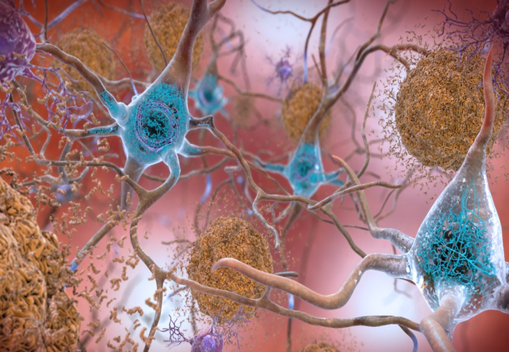
“Beta-Amyloid Plaques and Tau in the Brain” by national Institutes of Health (NIH) is marked with CC OM 1.0
Brain Development
Basic Brain Facts
At the cellular level, the brain is made up of 86 billion nerve cells called neurons that regulate cognitive activity. There are at least 10 times more support cells, called glial cells. Once it was thought we had 100 billion nerve cells but newer research has demonstrated it is actually 86 billion (BrainFacts/SFN, 2018). Neurons communicate with each other through billions of connections in an electrochemical process. There are about 500 trillion connections in the adult human brain.
Neurons and their connecting fibers are extremely small. One-hundred billion connective fibers can fit in a space the size of a match head. One inch cube of cortical mass contains over 10,000 miles of connective tissue.
Although there has long been a debate about whether we are more impacted by nurture (our environment) or nature (our individual biology), we now understand that it is actually a unique combination of both. While neither nature nor nurture fully explains what makes us human, we do know that it is a complex relationship between the two. Biology and genetics may provide the potential, but our social environment can shape our ability to access that potential.
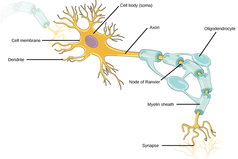
A neuron is made up of a dendrite, cell body and axon. Between neurons there is a small gap called the synaptic gap.
A dendrite and its spines receive information from other neurons. The number of dendrites on a neuron varies from a few hundred to thousands. Dendrites are covered with spines (varicosities) that are neurotransmitter receptor sites.
The cell bodyand its DNA genetic system use the nutrients that the blood brings to maintain the cell and to synthesize neurotransmitter molecules (messengers between cells).
The axonsends information from the neuron to other neurons. Each neuron generally has one axon branching out into many terminals. Axons vary in length from 1 millimeter to about 3 feet! Mature axons are covered in an insulated coating, which looks like sausage links, called myelin.
The synaptic gap is the tiny space between neurons, the neurons don’t actually touch. Neurotransmitters are released into the gap that act as chemical messengers to the receiving neuron.
Neurons transmit information between each other through axons and dendrites using the synaptic gap to exchange neurotransmitters. The axon sends a message through a series of electrical impulses called the action potential. When the impulse reaches the end of the axon the electrical activity ceases. A chemical process takes place in the form of neurotransmission. If the message is “transmit” an electrical charge is triggered in the next neuron. That neuron’s dendrite receives the message and electrically sends it through the axon to the next neuron. The process repeats until the message has reached its destination.
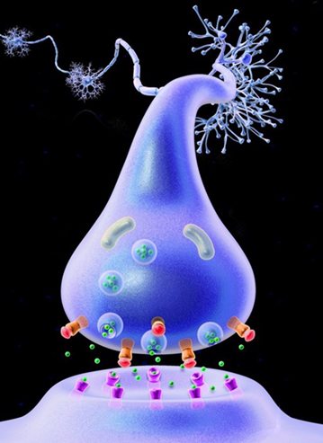
Neurotransmission
When the electrical impulse that carries information reaches the end of a neuron’s axon, they are stopped at the tiny synaptic gap that separates them from the receiving neuron. The circuit is broken. Neurotransmitters are chemical messengers secreted at the synapse that have the potential to continue the circuit and transmitting information between neurons.
Without neurotransmitters the brain could not process information or send out instructions to run the rest of the body. They affect the formation, maintenance, activity and longevity of synapses and neurons. Neurotransmitter molecules are produced within a specific type of neuron (different neurons are specialized in different neurotransmitters) and stored in tiny sacs known as vesicles. When an electrical signal reaches the vesicles, they release their neurotransmitters into the synaptic gap.
Each type of neurotransmitter has a unique shape that acts like a key. Released neurotransmitters attempt to attach to receptor sites (usually on the receiving neuron’s dendrites). Each receptor site is shaped like a lock that will fit only certain types of neurotransmitters. If the key fits, the neurotransmitter will send a message to turn on a receiving neuron- excitatory or off – inhibitory.
When a neurotransmitter’s job is done, the receptors release the molecules, which are either broken down or recycled. Each neurotransmitter has a very specialized function. Ligands or neurotransmitters can be broken down into categories: classical neurotransmitters, peptides, soluble gases, or steroids (hormones). Some neurotransmitters carry emotional information that impact our mood, outlook on life and behavior. For example, Cortisol has an impact on our stress response system.
Babies are born with an estimated 86 billion brain cells. We create new connections, in the form of neural pathways, in response to our active engagement in stimulating experiences. In the first few years of life more than 1 million new neural connections form every second (Center for the Developing Child). Most neural pathways are created after birth as a result of stimuli coming from the environment that the child interacts with through the senses.
Each time the brain responds to a similar stimulus there is an increased propensity for the neurons to reconnect along the same pathway. Connections grow in a brain when experiences are repeated over and over or when an experience triggers a strong emotional reaction. The brain becomes hard wired to respond along established pathways.
Neurons physically change as a result of this activation. Neurons grow new dendrite branches and receptor sites allowing the brain to process information more effectively and efficiently in more areas of the brain. The brain changes in response to experience by making connections with new input to what is already known and in place. The brain learns by recognizing patterns to make sense of new experiences. For example, when a baby tracks a toy with their eyes while grasping at it with their hand their visual and motor pathways are connecting and growing stronger. Experience literally sculpts the brain!
The most active period for creating connections is in the early years of life, but new connections can form throughout life. After this rapid proliferation early on, unused brain cells and connections wither away in a process called pruning. Pruning is necessary in order to make room for the pathways the child needs most to survive in their world. Creating room also has the function of making the remaining pathways more efficient. Think of how pruning a fruit tree is essential to make room for new growth and fruit to mature. Pruning too many neurons that are important will decrease the brain’s efficiency. Most intensive pruning happens between the ages 7 and 12 but is happening in some form throughout life, starting at about 8 months. The intensity of the pruning is dependent on which area of the brain is being affected at the time.
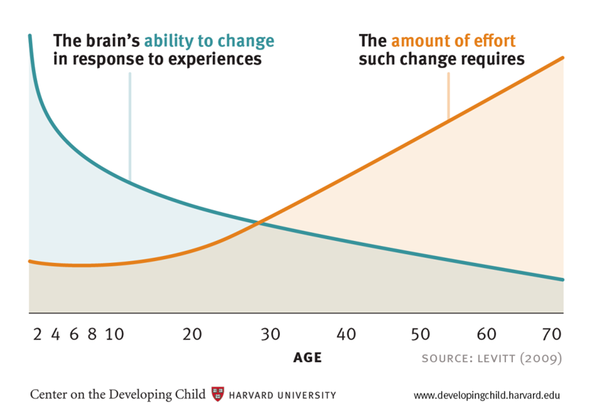
Plasticity
Plasticity is the term that describes the ease with which the brain can change itself. Our genes provide the blueprint, and our experiences are the architect. Which genes get turned on or off is determined by our experiences and environment. The brain’s pathways strengthen as they are used. As stated above, the neurons that are not used are subject to pruning, so it is a literal “use it or lose it” scenario. There is a remarkable increase in synapses during the first year of life. At birth we start with around 50 trillion connections, by 3 years we have around 1,000 trillion connections and as adults we have about 500 trillion connections. The brain is most plastic early in life and it is easier to influence a baby’s brain than try to rewire parts of it in the later years.
Image 4.x Rethinking the Brain is licensed under CC by 1.0
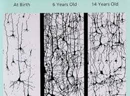
Picture of Synaptic Density: Synapses are created with astonishing speed in the first three years of life. For the rest of the first decade, children’s brains have twice as many synapses as adults’ brains (CHEF, 2003).
Windows of Opportunity
There are some windows of opportunity in the brain for optimal growth. During certain stages of brain growth, parts of the brain become much more active in response to what the senses absorb, growing and learning faster than at any other time in life. Children need the right experiences at the right time for their brains to fully develop in these areas. Sight is one of these windows of opportunity. If the eyes are deprived of sensory input early in life the neurons poised to connect for visual pathways reassign and sight will not develop. Most windows of opportunity are only optimal times and not absolutes. Every child is on their own timetable and so the age they reach the window will vary.
Table of 4 major developmental areas and windows of opportunity. CHEF 2003
Developmental Area:
Moving, Sensing and Exploring
Developmental Area:
Seeing, Remembering and Imagining
Developmental Area:
Listening, Talking and Understanding
Developmental Area:
Feeling and Relating
Windows of Opportunity:
Birth - 5 years: large motor
Birth - 10 years: fine motor
Windows of Opportunity:
Birth - 2 years: visual acuity
6 months - 13 years: visual memory
3-7 years: imagination
Windows of Opportunity:
prenatal - 3 years: first language
prenatal - 10 years: second language
1 year - 10 years: math and logic
3 - 10 years: music performance
Windows of Opportunity:
Prenatal - 1 year: trust
Prenatal - 18 months: secure attachments
birth - 3 years: self-esteem
8 months - 2 years: self-soothing
8 months - late adolescence: emotional skills
2 years - late adolescence: emotional control
It is important to remember that windows of opportunity are times when the brain is the most responsive for optimal development. When developmental stages are interrupted or skipped, or an injury of any degree is experienced, some sensory-motor and cognitive functions may be impaired or missing. For most functions it is never too late to grow new neurons and pathways, but it gets increasingly harder to do this as the brain ages. Early intervention is key to helping the brain get back on track for optimal development. The human brain has a remarkable ability to heal. (CHEF 2003) Windows don’t “slam shut” but slowly close as we age, never really shutting for good.
Children need active involvement in a stimulating, challenging and loving environment to cause the brain to grow and flourish. Passive involvement, isolation and an impoverished environment diminish the brain.
Enriched Environments
What is included in an enriched environment for the brain? Sleep: babies, children and adult’s brains need adequate sleep (see figure 1). Sleep is when the brain renews itself and cements learning. Nutrition: brains need proper nutrition with the right types of fat, protein, fruits and vegetables. We are quite literally what we eat, and our brain can only function as well as the fuel we give it. Foods high in refined sugar are toxic for a growing brain. The American Association of pediatrics recommends limiting the amount of sugar children consume each day to no more than 6 teaspoons for ages 2 and older. A typical child consumes more than triple that on average. (Jenco 2016) A great resource to make sure you are giving kids a balanced diet is MyPlate by the USDA. https://www.myplate.gov/eat-healthy/what-is-myplate
Water is also essential for the brain and body to stay hydrated. Encouraging children to drink water instead of juice is important to reduce the amount of sugar they are consuming while hydrating their brain. WebMD suggests the following: Toddlers need 2-4 cups, 4-8 years need 5 cups, 9-13 year need 7-8 cups and over 14 need 8-11 cups. (WebMD 2016)
Children need a safe environment with appropriate boundaries. Giving kids the freedom to explore while making sure that the environment is free from toxins and hazards helps young brains grow. They need the chance to interact with interesting materials and be given clear guidance about what is safe and not safe. We can think of boundaries as a fence we provide that surrounds the child and enlarges as they mature. The fence keeps them safe but within it they are free to explore and push against the boundary, so they know they are safe.
Another important part of an enriched environment is positive role models and guidance. Adults should model the lifestyle and behavior they want from children. Eating healthy, drinking water, getting adequate sleep and exercise, and modeling emotional intelligence and growth mind set skills are all part of this. If the adults around children strive to keep their brains healthy chances are kids will follow in suit. Positive guidance lets the child know they are safe, and that behavior is a learned skill just like tying their shoe. Both require activation of neurons to build strong pathways.
Young brains do best when media is limited, and they have daily exercise with time in nature. Movement of bodies creates an increase in the oxygen and blood flow to the brain, helping to keep it healthy at any age. Nature provides the brain with a complex bath of sensory input that will strengthen pathways and connections in a way that can’t be replicated indoors. In addition, our brains need down time and unstructured play. Down time for brains allows children to follow their own interests and develop mastery over skills they are learning. It is through unstructured play time that children feel free to learn about their world and strengthen their abilities. Young brains need practice repeating positive developmentally appropriate experiences with caring adults supporting them.
It is important not to stress the child by pushing them to do things they are not ready for or providing an overstimulating environment. The best approach is to follow the lead of the child and focus on their interests and unique timetables.
The child’s brain is not a smaller version of an adult brain. Neurons are still moving into position. As the brain develops, neurons migrate from the inner surface of the brain to form the outer layers. Immature neurons use fibers from cells called glia as highways to carry them to their destinations.
Figure 1: Recommended sleep by age group
Age Range
Recommended Hours of Sleep
Newborn
0-3 months old
14-17 hours *includes naps
Infant
4-11 months old
12-15 hours *includes naps
Toddler
1-2 years old
11-14 hours *includes naps
Preschool
3-5 years old
10-13 hours *includes naps
School-age
6-13 years old
9-11 hours
Teen
14-17 years old
8-10 hours
Young Adult
18-25 years old
7-9 hours
Adult
26-64 years old
7-9 hours
Older Adult
65 or more years old
7-8 hours
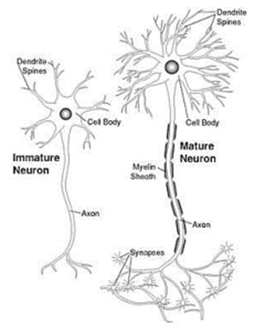
Myelination
Mature neurons have axons that are coated by a fatty layer called myelin, the protective sheath that covers communicating neurons. Myelin acts in two ways: it provides substance for the brain and insulates the cells. The myelination of axons speeds up the conduction of nerve impulses, through an ingenious mechanism that does not require large amounts of additional space or energy. Areas of the brain do not function efficiently until they are fully myelinated. Babies are born without much myelin.
Picture of immature neuron compared to a mature neuron. CHEF 2003
Myelin is composed of 30% protein and 70% fat. One of the most common fatty acids in myelin is oleic acid, which is also the most abundant fatty acid in human milk, and we should strive to include this in our diets. Monosaturated oleic acid is the main component of olive oil as well as the oils from almonds, pecans, macadamias, peanuts, and avocados. According to Harvard Health, how the brain begins is how it stays for the rest of life, so it is important to make sure nerves grow and connect and get covered with myelin. The essential nutrients for brain growth include:
Protein. Protein can be found in meat, poultry, seafood, beans and peas, eggs, soy products, nuts and seeds, as well as dairy.
Folate. This nutrient, which is especially important for pregnant mothers, can be found in liver, spinach, fortified cereals and breads, as well as other foods.
Iodine. Seaweed is a great source of iodine, but we also get it from iodized salt, seafood, dairy products, and enriched grains.
Vitamin D. This is the “sunshine vitamin,” and the best way to get it is to get outside. The flesh of fatty fishes such as salmon have it, as does fish liver oil, and products fortified with it, such as fortified milk. (McCarthy 2018)
In order to protect a babies’ unmyelinated neurons, it is important to never shake a baby. Although there may be no outside sign of damage the neurons get whipped around and have no protection from myelin. It is also essential that children get proper kinds and amounts of fats and oils. Breast milk contains a fat almost identical to the fat in myelin, so if possible, mothers should nurse during the first year of life.
The brain has boundaries around how quickly it can develop that are established by myelination timetables. Myelination can be stimulated when the brain is ready, but it cannot be rushed. Pushing a child to do something before they are ready can result in learning problems later on. Follow the child’s cues: their interest and frustration level will tell you when their brain is ready to learn a new skill.
Myelination continues to develop slowly all during childhood and adolescence in a gradual progression from lower to higher level systems. Early childhood is spent primarily on the brain stem, cerebellum and sensory cortex. Puberty is when the limbic system is primarily being myelinated and late adolescence the prefrontal cortex finishes myelination.
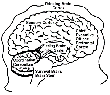
Layers of the Brain
The brain develops sequentially from the brainstem up, with the cortex developing last and continuously throughout life.
The Brain stem and midbrain are the first to develop and are mostly concerned with survival. The autonomic nervous system is regulated by the brain stem. It is the first part to mature. Babies are born with autonomic nervous system neurons fully myelinated. These neurons control survival needs such as heartbeat, breathing, and sucking. The brain stem and mid-brain monitor the outer world through sensory input and activate the body to respond in ways that ensure self-preservation.The brain stem processes information at a subconscious level; it is quick and reactive. Some of its functions include autonomic nervous system, fight/freeze/flight response, defense mechanisms, territoriality, reflexes, rote responses, routine, and habits. It is the least plastic layer of the brain and the most highly resistant to change. The reason habits are so hard to break is because they reside in this region of the brain. This part of the brain is often referred to as the “old brain” or “reptilian brain”.
Illustration by Victoria Tennant, Brain Child, V.T. Consulting, 2000
The Cerebellum is mostly in charge of coordination. It controls automatic movements and balance and the coordination of movement and thought or balance. The cerebellum is where procedural memory is stored like our motor skills. It does not involve conscious thought except when we are first learning something (like riding a bike). This area of the brain matures in early childhood and works in coordination with the brain stem.
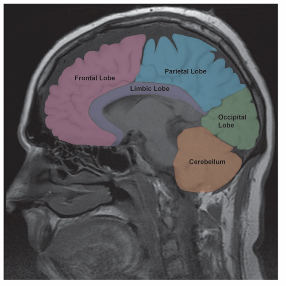
The limbic system is where emotions are processed. The limbic system is made up of many structures in the middle of the brain including the amygdala, hippocampus, thalamus, and olfactory bulbs. This area receives, interprets, and responds to emotional signals sent from the body. It processes information at the subconscious level and forms emotional patterns. This area is associated with long term memory and matures during puberty. It is considered the “heart” of the brain.
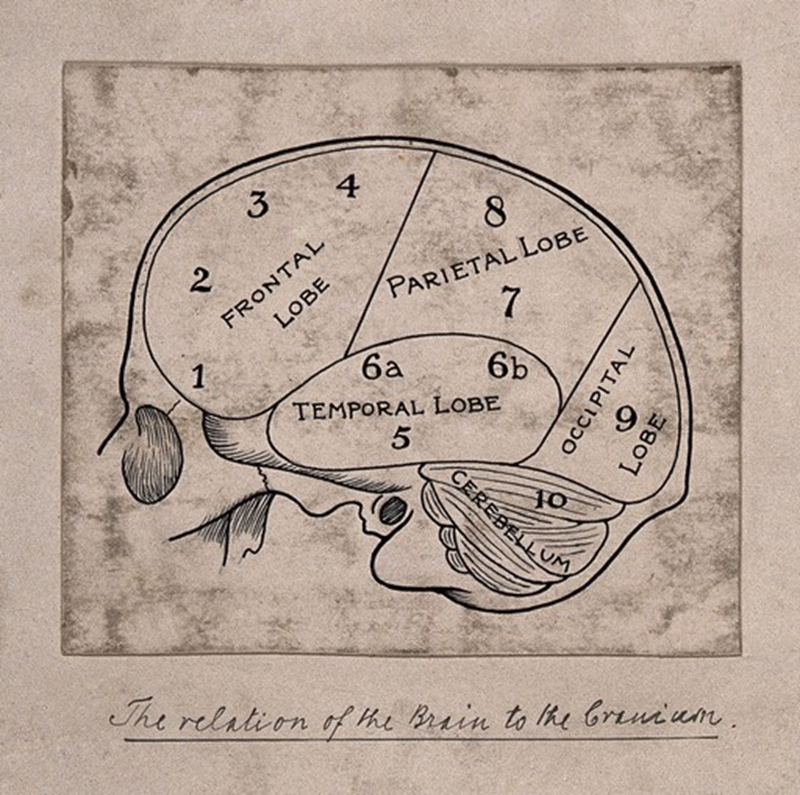
The cortex is where higher level thinking at the conscious level occurs. This includes, making sense of the world, decision making, creativity, reason, logic, imagination, self-awareness, and self-control. Everything that makes us uniquely human is the result of the interplay between the cortex working in harmony with the lower brain structures. The cortex loves change, novelty, fresh input and variety. It is the most plastic layer of the brain. The cortex is divided into specialized areas called lobes that are determined by their function. It matures over a long period of time, from the back to the front of the brain.
The cortex is split up into areas that are responsible for different functions. The back lobes are mostly related to sensory functions.
The occipital lobe is mainly responsible for vision and develops very early. The temporal lobe processes hearing, speech, language, and memory. The parietal lobe processes incoming sensory information like touch, pressure, pain, cold, heat, taste, and proprioception. The frontal lobe is responsible for gross and fine motor movements.
The prefrontal lobe, the very front section of the frontal lobe, is responsible for the critical thinking, creative thinking and problem solving. It is the part of the brain that allows us to imagine, plan and rehearse future actions. This area connects to the limbic system to regulate emotions. It is this integration of emotions with thought that is essential to the decision-making process. This area of the brain starts to develop around 8 moths and continues to develop late into adolescence (around age 26).
Executive Function and Self-regulation are also associated with this area of the brain. A child who develops the ability to self-regulate has better impulse control, mental flexibility, and emotional intelligence. These functions are critical for learning.
Although children do not have these skills from birth, they can be strengthened through practice with games and activities specifically aimed at reinforcing these skills. (See Activity 2).
“Growth-promoting environments provide children with “scaffolding” that helps them practice necessary skills before they must perform them alone. Adults can facilitate the development of a child’s executive function skills by establishing routines, modeling social behavior, and creating and maintaining supportive, reliable relationships. It is also important for children to exercise their developing skills through activities that foster creative play and social connection, teach them how to cope with stress, involve vigorous exercise, and over time, provide opportunities for directing their own actions with decreasing adult supervision.”