Up to this point, we’ve focused on the first two of the three problems associated with artifacts, namely noise and systematic differences in voltage across conditions. The remainder of the chapter will focus on the third problem, namely that blinks and eye movements can change the sensory input when visual stimuli are used.
For most researchers, this is not a big issue. Once you’ve dealt with the noise produced by blinks and eye movements (either by rejection or correction), problematic changes in sensory input are relatively rare. If you’re using artifact correction for blinks, you should still reject any trials with a blink that occurs at the time of the stimulus, because these trials are obviously not valid (see the chapter on artifact correction for details). And if the stimuli are presented in the middle of the display, participants won’t make a lot of eye movements, and if they do, they probably won’t differ systematically across conditions. This is what we saw in the MMN experiment. Some participants made eye movements as they watched the silent movie in the middle of the display, but these eye movements didn’t vary across conditions, and they couldn’t impact the sensory processing of the main stimuli (the auditory tones). They were a source of noise, but not a confound.
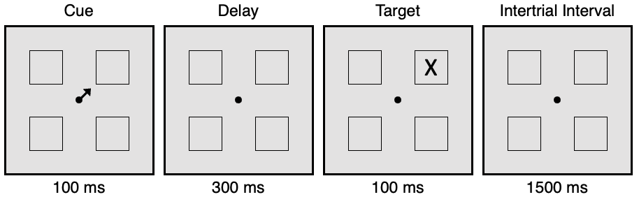
However, eye movements can be a significant systematic confound in some types of studies, mainly those using peripheral visual stimuli. For example, consider the spatial cuing paradigm shown in Figure 8.4, in which an arrow is used to indicate the likely location of a subsequent target. Many studies have used this paradigm to determine whether covert shifts of visual attention to the cued location cause the P1 and N1 waves to be larger when the stimulus is presented at the cued location compared to the uncued location (e.g., Eimer, 1994; Luck et al., 1994; Mangun & Hillyard, 1991). However, participants are likely to shift their gaze toward the cued location in these studies. If that happens, the target will appear in the center of gaze when it is presented at the cued location, whereas it will appear in the periphery when it is presented at an uncued location. We know that foveal stimuli produce larger sensory responses than peripheral stimuli, so this difference in the retinal location of the stimuli is a major confound that must be avoided in these studies. To avoid this confound, we can reject trials with eye movements. However, this is more difficult than it seems, even if you’re using a high-quality eye tracker. The next exercise will explain how to do this effectively.
Figure 8.4. Prototypical spatial cuing paradigm. The cue indicates the likely location of the target. After a short delay, the target appears at the cued location (80% of trials) or one of the uncued locations (20% of trials). Participants press one of two buttons, as quickly as possible, to indicate whether the target is an X or an O. Participants are also instructed to maintain fixation on the central point and focus their “covert” attention onto the cued location. The goal is to determine whether sensory processing is enhanced at the cued location relative to the uncued location.
Small eye movements are also a problem in studies that look at lateralized visual ERP components, such as the N2pc component and contralateral delay activity (CDA). Both of these components are negative voltages contralateral to the location of a to-be-perceived or to-be-remembered object or set of objects. There are two specific problems that arise in these experiments. First, if the eyes move to the relevant location, then this location is now foveal, and that may impact the lateralization that would otherwise be observed. This problem is especially acute if the stimulus is presented for more than 200 ms, which is the approximate amount of time required to make a controlled eye movement in these paradigms. With long stimulus durations, you may have one period of time in which the relevant stimuli are lateralized (prior to the eye movement) and then another period in which they are foveal (after the eye movement). Even with brief stimulus durations, however, changes in eye position could potentially change the lateralization of processing after the stimulus has disappeared (because the brain may remap the prior location of the internal neural representation onto its new retinal location).
The second problem is not due to the change in the sensory per se but is instead a confound in the EOG voltage. If participants tend to look leftward when the relevant stimuli are on the left side and rightward when the relevant stimuli are on the right side, then the EOG will be negative on the right side of the head when the relevant stimuli are on the left side and negative on the left side of the head when the relevant stimuli are on the right side. In other words, the EOG will appear as a negative voltage over the hemisphere contralateral to the relevant information, just like the N2pc and CDA. Moreover, the EOG is so large that even a small eye movement in the direction of the relevant information can produce a contralateral negativity that is as large or larger than the N2pc and CDA. The next exercise describes how to address both of these problems.
In theory, eye movements can also be a confound in studies of lateralized motor responses, such as the lateralized readiness potential (LRP; a negative voltage over the hemisphere contralateral to the response hand). This is because participants may make a small, unconscious eye movement toward the hand that responds. Such eye movements would produce a negative voltage over the right hemisphere for a left-hand response and a negative voltage over the left hemisphere for a right-hand response. That’s the same pattern as the LRP. The strategy described in the following sections for eliminating these small eye movements for the N2pc and CDA can also be used for the LRP.
If you don’t use lateralized visual stimuli or look at the LRP, then you can probably skip the rest of the chapter. However, you might want to read it and do the exercises anyway, because they provide good examples of the general principles of artifact rejection.