The psychophysiological methods discussed above focus on the central nervous system. Considerable research has also focused on the peripheral nervous system. These methods include skin conductance, cardiovascular responses, muscle activity, pupil diameter, eye blinks, and eye movements. Skin conductance, for example, measures the electrical conductance (the inverse of resistance) between two points on the skin, which varies with the level of moisture. Sweat glands are responsible for this moisture and are controlled by the sympathetic nervous system (SNS). Increases in skin conductance can be associated with changes in psychological activity. For example, studying skin conductance allows a researcher to investigate whether psychopaths react to fearful pictures in a normal way. Skin conductance provides relatively poor temporal resolution, with the entire response typically taking several seconds to emerge and resolve. However, it is an easy way to measure SNS response to a variety of stimuli.
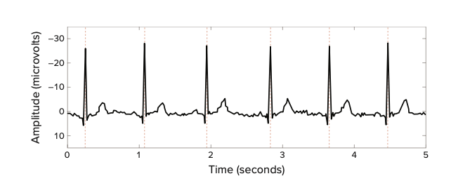
Cardiovascular measures include heart rate, heart rate variability, and blood pressure. The heart is innervated by the parasympathetic nervous system (PNS) and SNS. Input from the PNS decreases heart rate and contractile strength, whereas input from the SNS increases heart rate and contractile strength. Heart rate can easily be monitored using a minimum of two electrodes and is measured by counting the number of heartbeats in a given time period, such as one minute, or by assessing the time between successive heartbeats. Psychological activity can prompt increases and decreases in heart rate, often in less than a second, making heart rate a sensitive measure of cognition. Measures of heart rate variability are concerned with consistency in the time interval between heartbeats. Changes in heart rate variability are associated with stress as well as psychiatric conditions. Figure \(\PageIndex{1}\)is an example of an electrocardiogram, which is used to measure heart rate and heart rate variability. These cardio- vascular measures allow researchers to monitor SNS and PNS reactivity to various stimuli or situations. For example, when an arachnophobe views pictures of spiders, does their heart rate increase more than that of a person not afraid of spiders?
Figure \(\PageIndex{1}\): Example of electrocardiogram. The number of strong negative spikes in the output during a given period of time represents the heart rate, whereas the difference in the spacing between those strong negative spikes represents the heart rate variability. [This work, “ECG Output,” is licensed under CC BY-NC-SA 4.0 by Judy Schmitt. It is a derivative of “Figure 3” by Zachary Infantolino and Gregory A. Miller/Noba, which is licensed under CC BY-NC-SA 4.0.]
Electromyography (EMG) measures electrical activity produced by skeletal muscles. Similar to EEG, EMG measures the voltage between two points. This technique can be used to determine when a participant first initiates muscle activity to engage in a motor response to a stimulus or the degree to which a participant begins to engage in an incorrect response (such as pressing the wrong button), even if it is never visibly executed. It has also been used in emotion research to identify activity in muscles that are used to produce smiles and frowns. Using EMG, it is possible to detect very small facial movements that are not observable from looking at the face. The temporal resolution of EMG is similar to that of EEG and MEG.
Valuable information can also be gleaned from eye blinks, eye movements, and pupil diameter. Eye blinks are most often assessed using EMG electrodes placed just below the eyelid, but electrical activity associated directly with eye blinks or eye movements can be measured with electrodes placed on the face near the eyes, because there is voltage across the entire eyeball. Another option for the measurement of eye movement is a camera used to record video of an eye. This video method is particularly valuable when determination of absolute direction of gaze (not just change in direction of gaze) is of interest, such as when the eyes scan a picture. With the help of a calibration period in which a participant looks at multiple, known targets, eye position is then extracted from each video frame during the main task and compared with data from the calibration phase, allowing researchers to identify the sequence, direction, and duration of gaze fixations. For example, when viewing pleasant or unpleasant images, people spend different amounts of time looking at the most arousing parts. This, in turn, can vary as a function of psychopathology. Additionally, the diameter of a participant’s pupil can be measured and recorded over time from the video record. As with heart rate, pupil diameter is controlled by competing inputs from the SNS and PNS. Pupil diameter is commonly used as an index of mental effort when performing a task.