3.6: Cell Division
- Page ID
- 59011
\( \newcommand{\vecs}[1]{\overset { \scriptstyle \rightharpoonup} {\mathbf{#1}} } \)
\( \newcommand{\vecd}[1]{\overset{-\!-\!\rightharpoonup}{\vphantom{a}\smash {#1}}} \)
\( \newcommand{\id}{\mathrm{id}}\) \( \newcommand{\Span}{\mathrm{span}}\)
( \newcommand{\kernel}{\mathrm{null}\,}\) \( \newcommand{\range}{\mathrm{range}\,}\)
\( \newcommand{\RealPart}{\mathrm{Re}}\) \( \newcommand{\ImaginaryPart}{\mathrm{Im}}\)
\( \newcommand{\Argument}{\mathrm{Arg}}\) \( \newcommand{\norm}[1]{\| #1 \|}\)
\( \newcommand{\inner}[2]{\langle #1, #2 \rangle}\)
\( \newcommand{\Span}{\mathrm{span}}\)
\( \newcommand{\id}{\mathrm{id}}\)
\( \newcommand{\Span}{\mathrm{span}}\)
\( \newcommand{\kernel}{\mathrm{null}\,}\)
\( \newcommand{\range}{\mathrm{range}\,}\)
\( \newcommand{\RealPart}{\mathrm{Re}}\)
\( \newcommand{\ImaginaryPart}{\mathrm{Im}}\)
\( \newcommand{\Argument}{\mathrm{Arg}}\)
\( \newcommand{\norm}[1]{\| #1 \|}\)
\( \newcommand{\inner}[2]{\langle #1, #2 \rangle}\)
\( \newcommand{\Span}{\mathrm{span}}\) \( \newcommand{\AA}{\unicode[.8,0]{x212B}}\)
\( \newcommand{\vectorA}[1]{\vec{#1}} % arrow\)
\( \newcommand{\vectorAt}[1]{\vec{\text{#1}}} % arrow\)
\( \newcommand{\vectorB}[1]{\overset { \scriptstyle \rightharpoonup} {\mathbf{#1}} } \)
\( \newcommand{\vectorC}[1]{\textbf{#1}} \)
\( \newcommand{\vectorD}[1]{\overrightarrow{#1}} \)
\( \newcommand{\vectorDt}[1]{\overrightarrow{\text{#1}}} \)
\( \newcommand{\vectE}[1]{\overset{-\!-\!\rightharpoonup}{\vphantom{a}\smash{\mathbf {#1}}}} \)
\( \newcommand{\vecs}[1]{\overset { \scriptstyle \rightharpoonup} {\mathbf{#1}} } \)
\( \newcommand{\vecd}[1]{\overset{-\!-\!\rightharpoonup}{\vphantom{a}\smash {#1}}} \)
Mitosis is the process of nuclear division used in conjunction with cytokinesis to produce 2 identical daughter cells. This process is used for somatic (body) cells that have the full amount of chromosomes (e.g., 46). Cytokinesis is the actual separation of these two cells enclosed in their own cellular membranes. Unicellular organisms utilize this process of division in order to reproduce asexually. Prokaryotic organisms lack a nucleus, therefore they undergo a different process called binary fission. Multicellular eukaryotes undergo mitosis for repairing tissue and for growth.
The process of mitosis is only a short period of the lifespan of cells. Mitosis is traditionally divided into four stages: prophase, metaphase, anaphase and telophase. The actual events of mitosis are not discreet but occur in a continuous sequence—separation of mitosis into four stages is merely convenient for our discussion and organization. During these stages important cellular structures are synthesized and perform the mechanics of mitosis.
For example, in animal cells two microtubule organizing centers called centrioles replicate. The pairs of centrioles move apart and form an axis of proteinaceous microtubules between them called spindle fibers. These spindle fibers act as motors that pull at the centromeres of chromsomes and separate the sister chromatids into newly recognized chromosomes. The spindles also push against each other to stretch the cell in preparation of forming two new nuclei and separate cells. In animal cells, a contractile ring of actin fibers cinch together around the midline of the cell to coordinate cytokinesis. This cinching of the cell membrane creates a structure called the cleavage furrow. Eventually, the cinching of the membrane completely separates into two daughter cells. Both daughter cells have the same number of chromosomes (in humans, 46) as each other and the same number as the original cell. This is the diploid number (full number).

Cells in the human body have 46 chromosomes, including 22 pairs of autosomes and one pair of sex chromosomes (XX in females, XY in males). Because there are two sets of chromosomes, one from each parent, the cells are considered diploid. Meiosis starts with a diploid cell and turns it into four haploid cells, cells with only one set of chromosomes. This means that when the chromosomes of egg and sperm cells combine at fertilization, the embryo regains the normal diploid number.
Meiosis mixes up the parental genes in two ways. First, the members of each chromosome pair come together and swap segments in a process known as crossing over, or recombination (see below). Second, because each gamete gets only half the parental chromosomes, the exact combination in each egg or sperm can and does vary. This is because during meiosis the chromosomes assort independently, with a random member of each pair going to each daughter cell.
Because males have one X and one Y chromosome, half the cells get an X and half get a Y during the meiosis that leads to sperm production. (In females, all the eggs will get one or the other X.) In a general sense, the sex of the offspring is determined by the particular sex chromosome carried by the sperm. However, in the early weeks of development, all fetuses have preliminary structures for both sexes, and the immature gonads can become either testes or ovaries. In the seventh week of fetal development, a gene on the Y chromosome, if present, activates, and the bipotential gonads commit to becoming testes. In the absence of a Y chromosome, and the signal to form testes, the fetus develops as a girl.
At least that's the way it usually happens. In rare cases, an XX individual becomes a male or an XY individual becomes female. Researchers realized that studying the genes of these sex-reversed people could lead them to the master switch for sex determination. They subsequently identified a gene called SRY (sex-determining region on the Y chromosome).
Meiosis, the form of cell division unique to egg and sperm production, sets the stage for sex determination by creating sperm that carry either an X or a Y sex chromosome. But what is it about the X or Y that determines sex? Before a meiotic cell divides, its two sets of chromosomes come together and cross over, or swap, segments. The first animation shows normal crossing over, where the X and Y chromosomes exchange pieces only at their tips. The second animation shows a rare mistake in which the Y chromosome transfers a gene called SRY to the X chromosome, resulting in sex-reversed babies. Studies of sex-reversed individuals led researchers to identify the master switch for sex determination, the SRY gene, which tells a fetus to become a boy.
What is different about meiosis is that there are two divisions instead of just one, as in mitosis. This is to make sure gametes (sex cells; sperm or eggs) only have the haploid amount (or half; e.g., 23) of chromosomes since each parent can only pass down half their genetic material.
Meiosis I
During the first meiotic division, recombination occurs and the chromosome number is halved.
Prophase I: Chromosomes condense and become visible. Homologous chromosomes pair up and recombination (crossing over) occurs. Crossovers may be visible as chiasmata, x-shaped connections between chromatids.
Metaphase I: Paired chromosomes line up along the cell's equatorial plane.
Anaphase I: Homologous pairs separate and move to opposite poles.
Telophase I: Chromosomes are at poles; nuclear membranes may re-form.

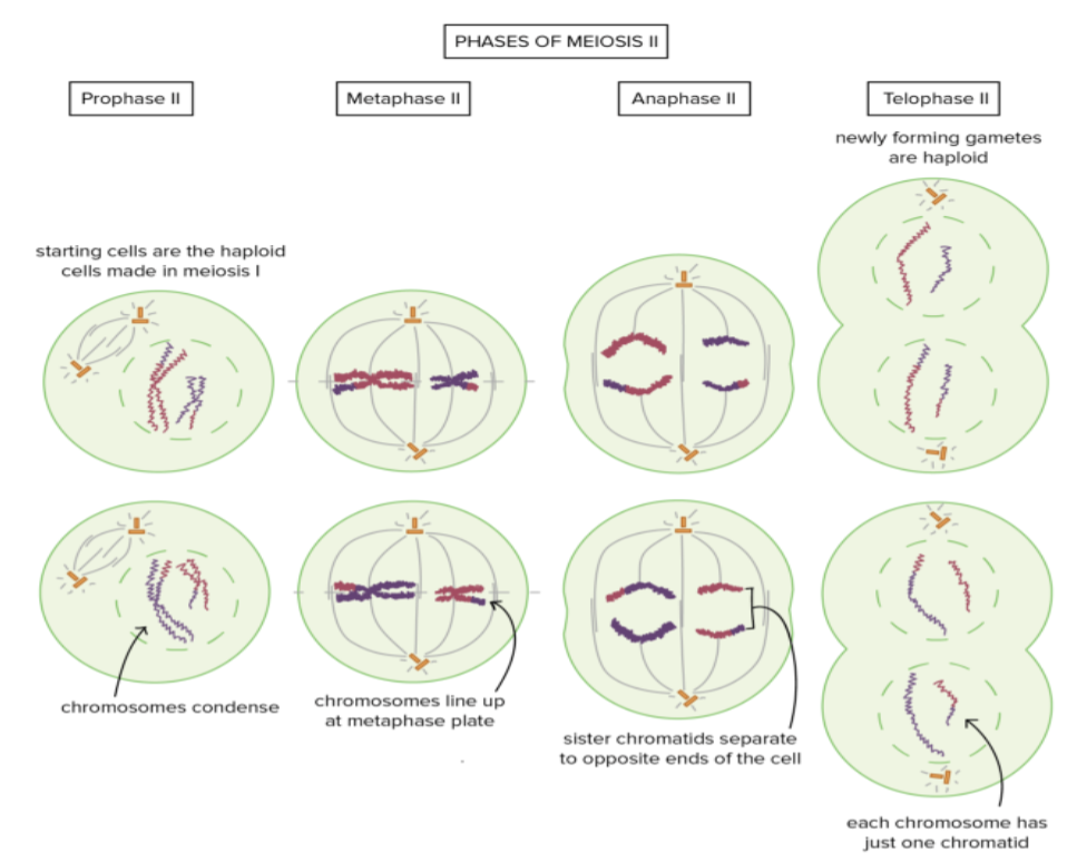
Meiosis II
The second meiotic division closely resembles mitosis (the type of cell division that occurs in body cells), except that the starting and ending cells are haploid.
Prophase II, metaphase II, anaphase II: The chromosomes again move to the equatorial plane, and this time the chromatids separate to opposite poles.
Telophase II: Nuclear membranes re-form around the chromosomes.