Osteology, or the study of bones, is central to biological anthropology because every person’s skeleton tells a story of how that person has lived. Bones from archaeological sites can be used to understand what animals people ate, how stressful and strenuous their lives were, and how they died—by natural or unnatural causes. This appendix introduces the basics of anatomical terminology and describes the different regions and bones of the skeleton. It also highlights some skeletal features that are used frequently by forensic anthropologists to estimate the age and sex of recovered remains. The authors note that sex is not binary but exists on a spectrum based on influences of chromosomes, genes, and hormones. These biological influences affect the size and shape of bone, which is sometimes useful in classifying skeletal remains into one of the two most common sex categories: female and male.
Bone Structure and Function
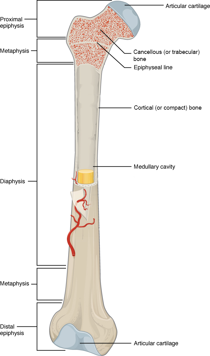
Bone is a composite of organic collagen and an inorganic mineral (hydroxyapatite, a calcium phosphate salt), which help make it strong enough to support the body under the force of gravity without collapsing. When bone is mature (fully mineralized as in adults), it comprises an outer dense region of bone called cortical (or compact) bone and an inner spongy region of bone called cancellous (or trabecular) bone (Figure A.1).
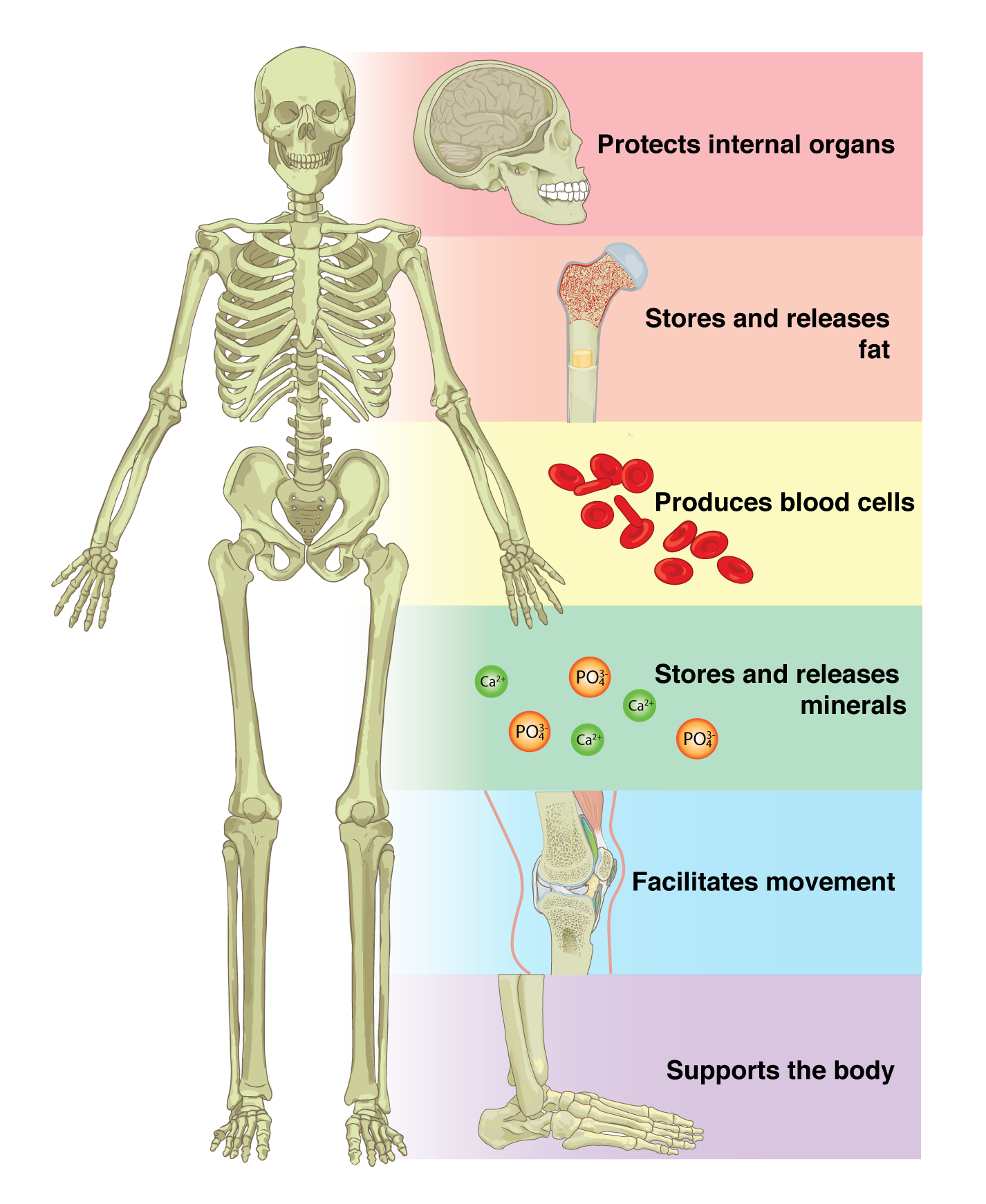
Bone performs both metabolic and mechanical functions for the body. On the metabolic side, bone is required to maintain mineral (i.e., calcium) homeostasis and for the production of red and white blood cells (Figure A.2), which develop in the diaphyseal marrow cavity and the cancellous region of the metaphysis and epiphysis. But it is undeniable that the mechanical functions of bone are primary because bone is critically responsible for protecting internal organs, providing support against the force of gravity, and serving as a network of rigid levers for muscles to act upon during movement.
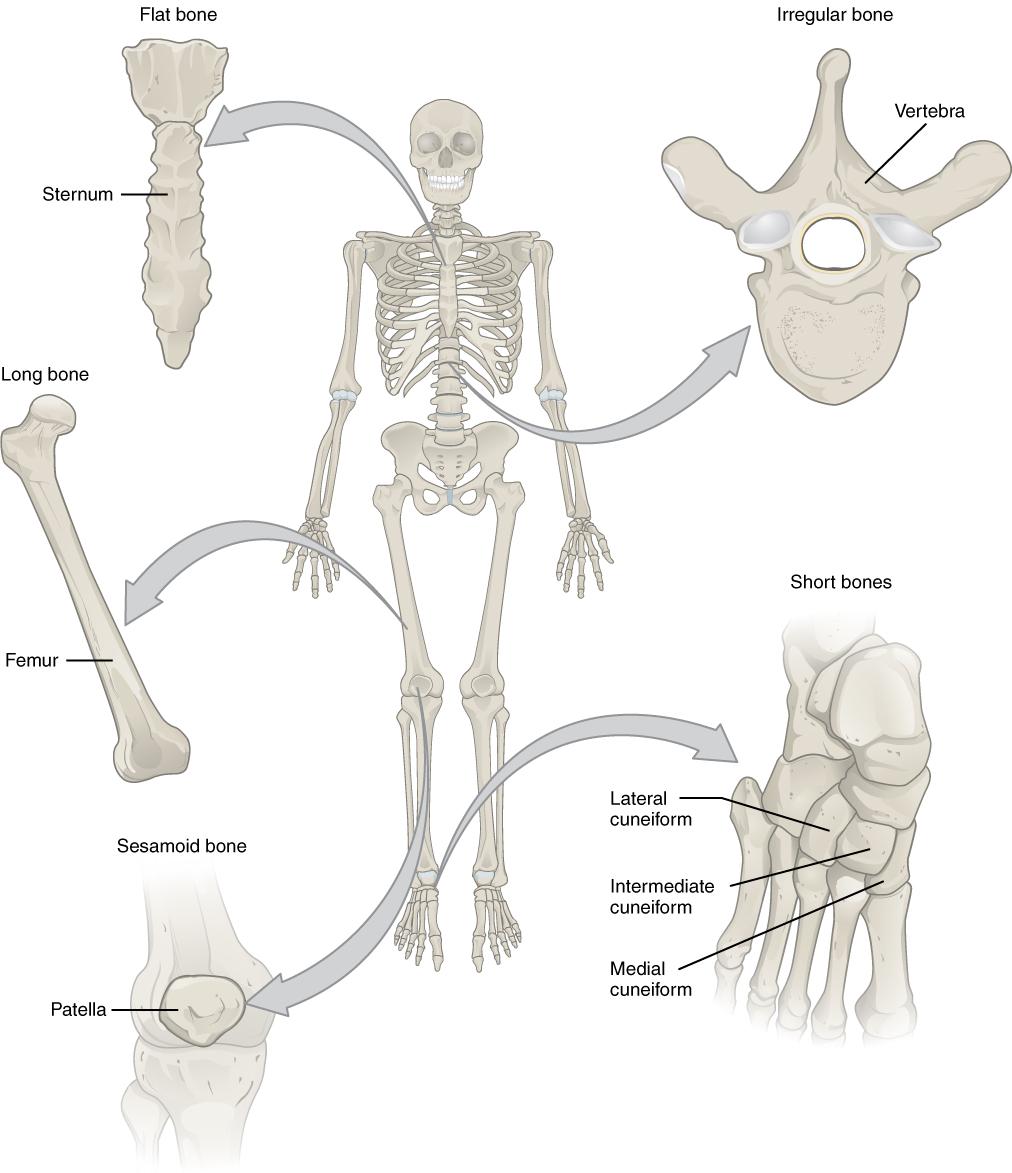
Bones have different shapes that largely relate to their specific function within the skeletal system. Additionally, the ratio of cortical to cancellous bone, and which muscles are attached to the bone and how, affect the shape of the whole bone. Generally there are five recognized bone shapes: long bones, short bones, flat bones, sesamoid bones, and irregular bones. Long bones are longer than they are wide and consist of three sections: diaphysis, epiphysis, and metaphysis (see Figure A.1). The diaphysis of a long bone is simply the shaft of the bone, and it comprises mostly cortical bone with a thin veneer of internal cancellous bone lining a medullary cavity. At both the proximal and distal ends of every long bone, there is an epiphysis, which consists of a thin shell of cortical bone surrounding a high concentration of cancellous bone. The epiphysis is usually coated with cartilage to facilitate joint articulation with other bones. The junction between diaphysis and epiphysis is the metaphysis, which has a more equal ratio of cortical to cancellous bone. Examples of long bones are the humerus, the femur, and the metacarpals and metatarsals.
The other three bone shapes are simpler. Shortbones are defined as being equal in length and width, and they possess a mix of cortical and cancellous bone (Figure A.3). They are usually involved in forming movable joints with adjacent bones and therefore often have surfaces covered with cartilage. Examples of short bones are the carpals of the wrist and the tarsals of the ankle. Flatbones are flat and consist of two layers of thick cortical bone with an intermediate layer of cancellous bone referred to as diploë. Most of the bones of the skull are flat bones, such as the frontal and parietal bones, as well as all parts of the sternum (Figure A.3). Sometimes bones develop within the tendon of a muscle in order to reduce friction on the joint surface and to increase leverage of the muscle to move a joint. These types of bones are called sesamoid bones, and these include the patella (or knee cap) and the pisiform (a bone of the wrist). Irregularbones are bones that don’t fit into any of the other four categories. The shapes of these bones are often more complex than the others, and examples include the vertebrae and certain bones of the skull, like the ethmoid and sphenoid bones (Figure A.3).
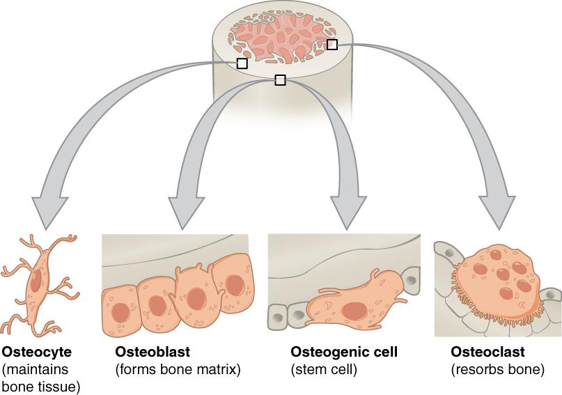
Each time we move our muscles, we bend, twist, compress, and tense our bones, and this causes them to develop microscopic cracks that weaken them. These may even lead to a bone fracture. Bone cells called osteocytes can sense when these microcracks form. Osteocytes then signal osteoclasts to remove the cracked bone and osteoblasts to lay down new bone—a process known as skeletal remodeling. Osteogenic cells are stem cells that are able to differentiate into osteoblasts and osteocytes (Figure A.4).
Figure A.4: Four types of cells are found within bone tissue. Osteogenic cells are stem cells that develop into osteoblasts. Osteoblasts lay down new bone while osteoclasts remove bone. Osteoblasts that get trapped in calcified matrix become osteocytes. Credit: Bone Cells (Anatomy & Physiology, Figure 6.11) by OpenStax is under a CC BY 4.0 License. [Image Description]
Did Deeper: How Do Bones Develop?
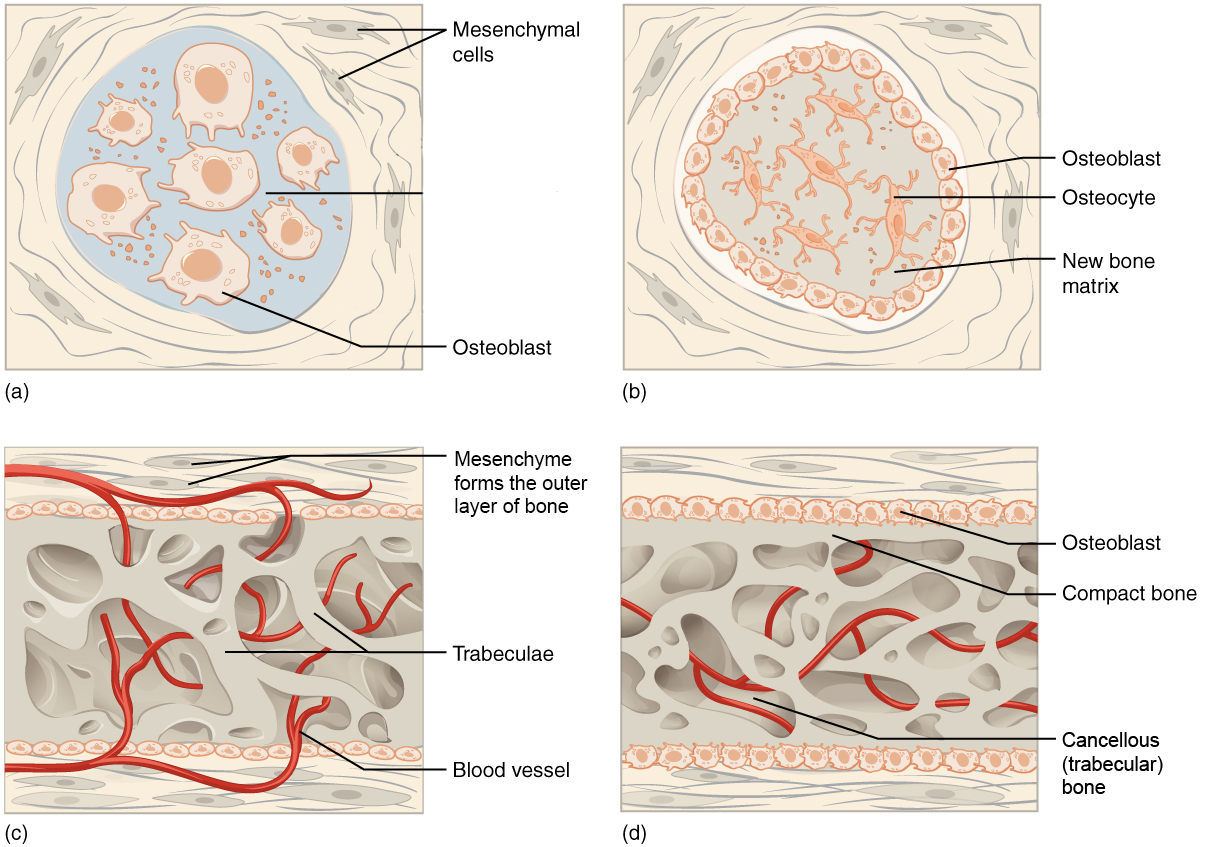
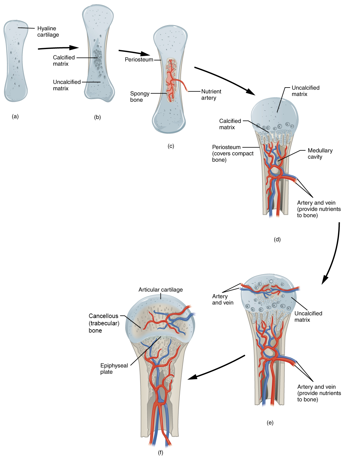
Bones develop via one of two mechanisms: intramembranous or endochondral bone formation. During intramembranous bone formation connective tissue (mesenchymal) stem cells form a tissue layer and then differentiate into osteoblasts, which begin to synthesize new bone along the tissue layer (Figure A.5). Only a few bones develop through intramembranous bone formation, mostly bones of the skull and the clavicle (collar bone). In endochondral bone formation, instead of developing directly from connective tissue stem cells, osteoblasts develop from an intermediate cartilage “model” that is then replaced by synthesized new bone (Figure A.6). Most bones of the skeleton develop through endochondral bone formation (Burr and Organ 2017).
Figure A.5: Intramembranous ossification begins when mesenchymal stem cells group into clusters. These clusters contain osteoblasts, which lay down the initial trabecular bone. Compact bone develops superficial to the trabecular bone, and the initial structure of the bone is complete. Credit: Intramembranous Ossification (Anatomy & Physiology, Figure 6.16) by OpenStax has been modified (some labels modified or removed) and is under a CC BY 4.0 License. [Image Description]Figure A.6: Endochondral ossification begins when mesenchymal cells differentiate into cartilage cells, which lay down a cartilage model of the future bony skeleton. Cartilage is then replaced by bone, except at the (epiphyseal) growth plates (which fuse at the end of postnatal growth) and the hyaline (articular) cartilage on the joint surface. Credit: Endochondral Ossification (Anatomy & Physiology, Figure 6.17) by OpenStax has been modified (some labels modified or removed) and is under a CC BY 4.0 License. [Image Description]