
3.2: The Peripheral Nervous System
- Page ID
- 170812
This page is a draft and under active development. Please forward any questions, comments, and/or feedback to the ASCCC OERI (oeri@asccc.org).
- Describe the structures found in the PNS
- Distinguish between somatic and autonomic structures
- Explain the functions of the sympathetic and parasympathetic divisions of the autonomic system
Overview
This module discusses the peripheral nervous system (PNS) in greater depth, including nervous tissues, the two major systems the PNS divides into (the somatic nervous system and the autonomic nervous system), names and functions of the twelve cranial nerves, and characteristics of the 31 pairs of spinal nerves. The subdivisions of the autonomic nervous system (the sympathetic nervous system and the parasympathetic nervous system) and their functions are also covered, as well as a brief mention of disorders of the PNS.
What Is the Peripheral Nervous System?
The peripheral nervous system (PNS) consists of all the nervous tissue that lies outside of the central nervous system (CNS). The main function of the PNS is to connect the CNS to the rest of the organism. It serves as a communication relay, going back and forth between the CNS and muscles, organs, and glands throughout the body. Figure \(\PageIndex{1}\) illustrates both the central and peripheral nervous systems, including many representative spinal nerves. Although an overview of spinal nerves is discussed in greater detail below, the identification and functions of specific spinal nerves are beyond the scope of this section.
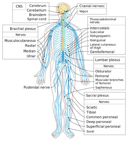
The representative nerves pictured are Cranial nerves: Vagus. Brachial plexus nerves: Musculotaneous, Radial, Median, Ulnar; Thoracoabdominal nerves: Intercostals, Subcostal, Iliohypogastric, Ilioinguinal, Lateral cutaneous of thigh, Genitofemoral; Lumbar plexus nerves: Obturator, Femoral, Muscular branches of femoral, Saphenous; Sacral plexus nerves: Sciatic, Tibial, Common peroneal, Deep peroneal, Superficial peroneal, Sural. The Pudendal nerve is also pictured.
Tissues of the Peripheral Nervous System
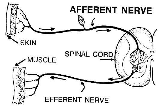
The primary structures that make up the PNS are nerves and ganglia. Ganglia are clusters of neuron cell bodies outside the CNS; they act as relay points for messages transmitted through nerves of the PNS. Nerves are cable-like bundles of axons that make up the majority of PNS tissues. Nerves are generally classified based on the type of information they carry (which coincides with the direction in which their nerve impulses travel) as sensory, motor, or mixed nerves. See an example of a sensory and motor nerve in Figure \(\PageIndex{2}\). (Note that the interneuron that is usually located between the sensory and motor neuron is not shown in this figure.)
- Sensory nerves transmit information from sensory receptors in the body to the CNS. Sensory nerves are also called afferent nerves.
- Motor nerves transmit information from the CNS to muscles, organs, and glands. Motor nerves are also called efferent nerves.
- Mixed nerves contain axons from both sensory and motor neurons, so they transmit information in both directions and have both afferent and efferent functions.
Divisions of the Peripheral Nervous System
The PNS is divided into two major systems, called the autonomic nervous system and the somatic nervous system. Both systems of the PNS interact with the CNS and include sensory and motor neurons, but they use different circuits of nerves and ganglia.
Somatic Nervous System
The somatic nervous system primarily senses the external environment and controls voluntary activities for which decisions and commands come from the cerebral cortex of the brain. For example, when you feel too cold, decide to turn on the heater, and walk across the room to the thermostat, you are using your somatic nervous system. In general, the somatic nervous system is responsible for all of your conscious perceptions of the outside world, and all of the voluntary motor activities you perform in response. Whether it’s playing piano, driving a car, or playing basketball, you can thank your somatic nervous system for making it possible. Structurally, the somatic nervous system consists of 12 pairs of cranial nerves and 31 pairs of spinal nerves.
Autonomic Nervous System
The autonomic nervous system primarily senses the internal environment and controls involuntary activities. It is responsible for monitoring conditions in the internal environment and bringing about appropriate changes in them. In general, the autonomic nervous system is responsible for all the activities that go on inside your body without your conscious awareness or voluntary participation.
Structurally, the autonomic nervous system consists of sensory and motor nerves that run between the CNS (especially the hypothalamus in the brain) and internal organs (such as the heart, lungs, and digestive organs) and glands (such as the pancreas and sweat glands). Sensory neurons in the autonomic system detect internal body conditions and send messages to the brain. Motor nerves in the autonomic system function by controlling the contractions of smooth or cardiac muscle or glandular tissue. For example, when sensory nerves of the autonomic system detect a rise in body temperature, motor nerves signal both the smooth muscles in blood vessels near the body surface to undergo vasodilation, and the sweat glands in the skin to secrete more sweat, to cool the body.
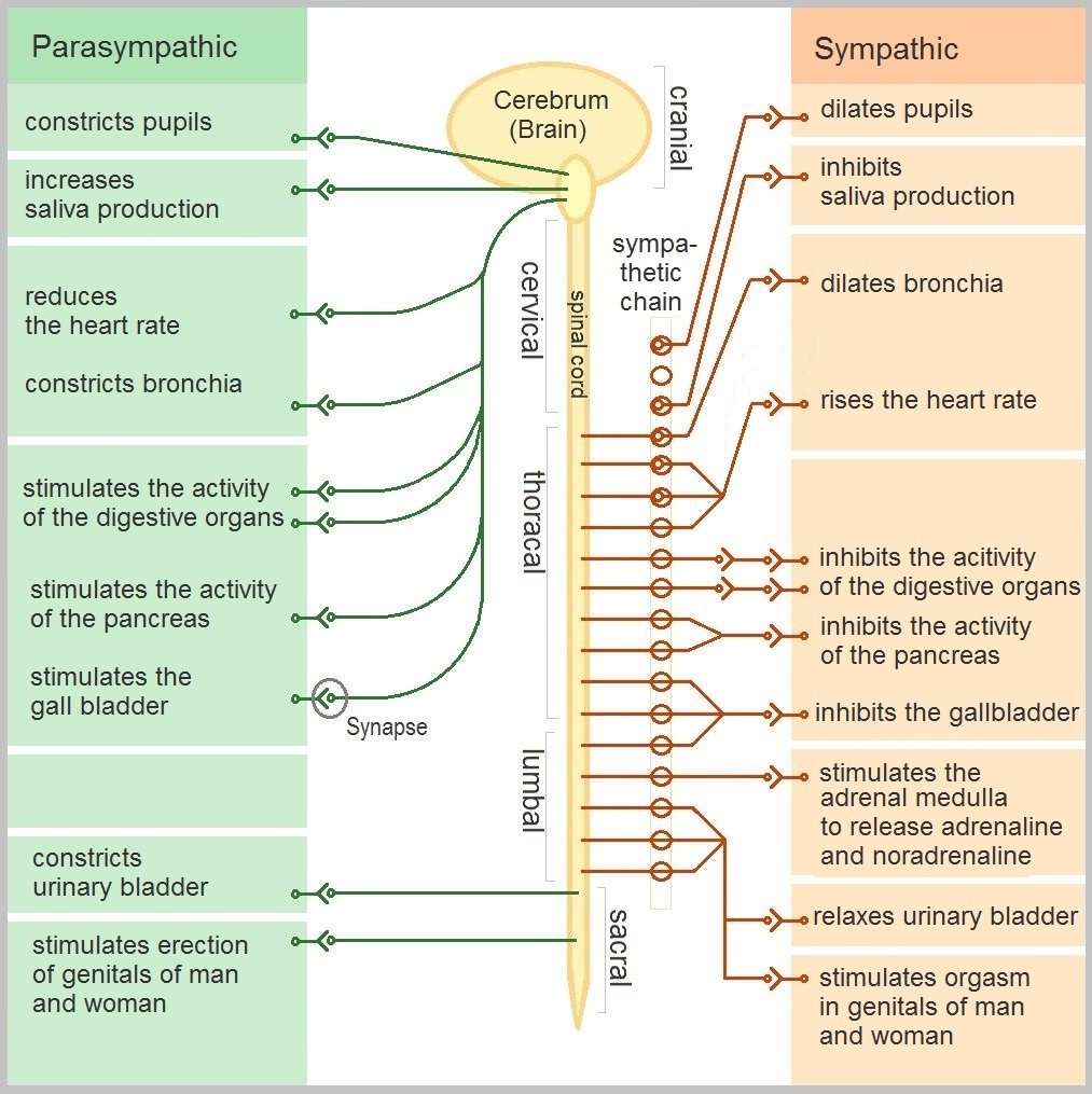
The autonomic nervous system, in turn, has two subdivisions: the sympathetic division and parasympathetic division. The functions of the two subdivisions of the autonomic system are summarized in Figure \(\PageIndex{5}\). Both affect most of the same organs and glands, but they generally do so in opposite ways.
- The sympathetic division controls the fight-or-flight response. Changes occur in organs and glands throughout the body that prepare the body to fight or flee in response to a perceived danger. For example, the pupils dilate, saliva production decreases, air passages in the lungs become wider (the bronchia dilate), heart rate speeds up, more blood flows to the skeletal muscles, and the digestive system temporarily shuts down. With regard to urinary and sexual functions, the sympathetic division relaxes the urinary bladder and stimulates orgasm in both men and women.
- The parasympathetic division returns the body to normal after the fight-or-flight response has occurred. For example, pupils constrict, saliva production increases, air passages in the lungs narrow (the bronchia constrict), heart rate slows down, blood flow to the skeletal muscles is reduced, and the digestive system is stimulated to start working again. The parasympathetic division also maintains the internal homeostasis of the body at other times. With regard to urinary and sexual functions, the parasympathetic division constricts the urinary bladder and stimulates genital tissue erection in both men and women.
Disorders of the Peripheral Nervous System
Unlike the CNS, which is protected by bones, meninges, and cerebrospinal fluid (CSF), the PNS has no such protections. The PNS also has no blood-brain barrier to protect it from toxins and pathogens in the blood. Therefore, the PNS is more subject to injury and disease than is the CNS. Causes of nerve injury include diabetes, infectious diseases such as shingles, and poisoning by toxins such as heavy metals. Disorders of the PNS often have symptoms such as loss of feeling, tingling, burning sensations, or muscle weakness. If a traumatic injury results in a nerve being transacted (cut all the way through), it may regenerate, but this is a very slow process and may take many months.
Summary
The peripheral nervous system (PNS) is composed of the groups of neurons (ganglia) and bundles of axons (nerves) that are outside of the brain and spinal cord. The PNS connects the CNS to the rest of the body.
Sensory (/afferent) nerves transmit information from sensory receptors in the body to the CNS. Motor (/efferent) transmit information from the CNS to muscles, organs, and glands. Mixed nerves transmit information in both directions and have both sensory (/afferent) and motor (/efferent functions). The PNS is divided into the autonomic nervous system and the somatic nervous system.
The autonomic nervous system senses the internal environment and controls involuntary activities. It is divided into the sympathetic division and parasympathetic division. Both affect most of the same organs and glands, but in opposite ways. The sympathetic division controls the fight-or-flight response and the parasympathetic division returns the body to normal after the fight-or-flight response has occurred.
The PNS does not have the same protections (bones, meninges, CSF, blood-brain barrier) that the CNS has, so it is more prone to injury and disease. If a nerve is transacted, it may regenerate, but this is a very slow process.
Additional Resources
Interested in the effects of meditation on the brain? See Integrative and Contemplative Neuroscience
Mindfulness techniques have been shown to reduce symptoms of depression as well as those of anxiety and stress. They have also been shown to be useful for pain management and performance enhancement. Specific mindfulness programs include Mindfulness-Based Stress Reduction (MBSR) and Mindfulness Mind-Fitness Training (MMFT). You can learn more about MBSR by watching the video below.
Ever wonder why "hot" peppers are perceived as hot? Check out this link:
Attributions
- Figures:
- The nervous system licensed CC BY-SA 4.0 via Lumen Learning
- Afferent nerve by Pearson Scott Foresman, Public domain via Wikimedia Commons
- Spinal Cord Segments and body representation by David Nascari and Alan Sved, CC BY-SA 4.0 via Wikimedia Commons
- Brain Spinal Cord Labeled by Vankadara Bhavya sree 1840585, CC BY-SA 4.0 via Wikimedia Commons
- Autonomic nervous system by Geo-Science-International, dedicated CC0 via Wikimedia Commons
- Text adapted from:
- " Peripheral Nervous System" by Suzanne Wakim & Mandeep Grewal, LibreTexts is licensed under CC BY-NC
- " The Peripheral Nervous System" by OpenStax, LibreTexts is licensed under CC BY . (Table also from this source.)
- Changes: Text (and images) from above four sources pieced together with some modifications, transitions and additional content (and images) added by Naomi I. Gribneau Bahm, PhD., Psychology Professor at Cosumnes River College, Sacramento, CA.