
3.3: The Central Nervous System
- Page ID
- 170813
This page is a draft and under active development. Please forward any questions, comments, and/or feedback to the ASCCC OERI (oeri@asccc.org).
- Name the structures that comprise the central nervous system
- Describe the structure and function of the spinal cord
Overview
This module defines the central nervous system (the brain and spinal cord).
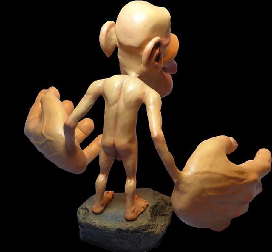
Figure \(\PageIndex{1}\) is a very odd-looking model called a homunculus. This model shows the relative representation of different parts of the body in the primary sensory cortex, an area in the parietal lobe of the brain. As you can see, larger areas of the brain in this region are associated with the hands, face, and tongue than the arms, torso, legs and feet. Larger areas of representation in the brain result in greater sensitivity in those body areas. Given the importance of speech, manual dexterity, and face-to-face social interactions in human beings, it is not surprising that relatively large areas of the brain are needed to relay sensation from these body parts. (There is a similarly odd-looking representation of voluntary motor functions called the motor homunculus that is located in the primary motor cortex, an area in the frontal lobe of the brain.) The brain is the most complex organ in the human body and is part of the central nervous system.
What Is the Central Nervous System?
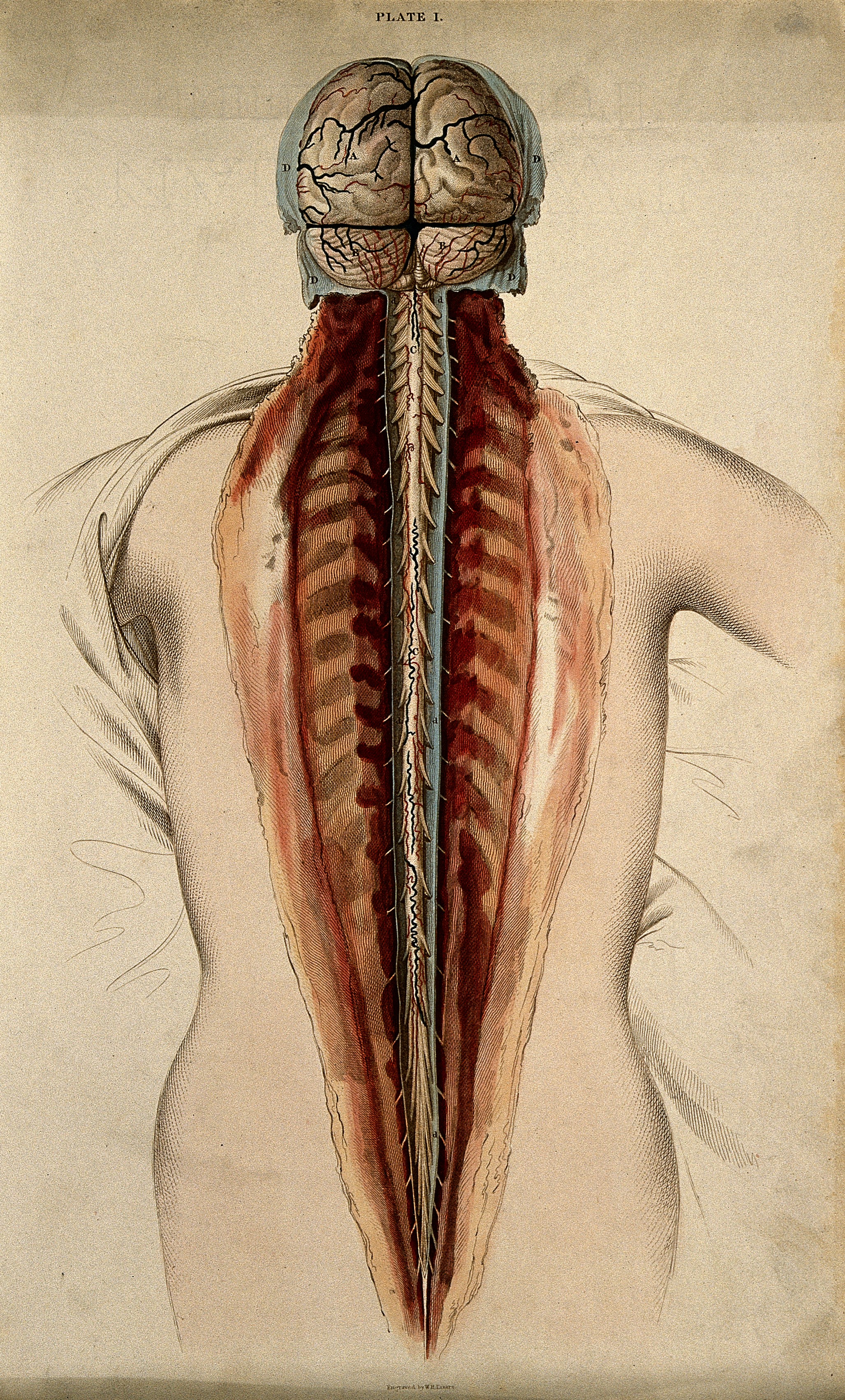
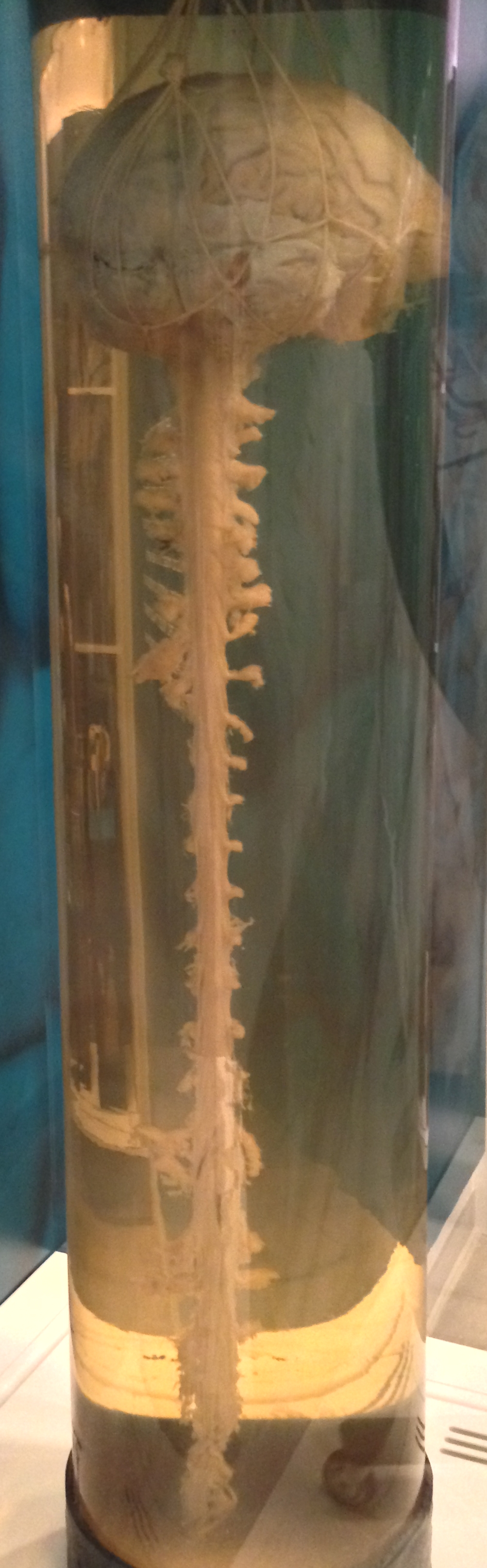
The central nervous system (CNS) is the part of the nervous system that includes the brain and spinal cord. Figure \(\PageIndex{2}\) shows the central nervous system both as it appears in the body dissected from the backside and also completely removed from the body. The central nervous sytem and the peripheral nervous system (PNS, the other main division, which is addressed in a separate module), work together to control virtually all body functions.
The Brain
The brain is the control center not only of the rest of the nervous system but of the entire organism. The adult brain makes up only about two percent of the body’s weight, but it uses about 20 percent of the body’s total energy. The brain contains an estimated 86 billion neurons (Lent et al., 2012), and each neuron has thousands of synaptic connections to other neurons. It is estimated that the brain also has about the same number of glial cells as neurons. No wonder the brain uses so much energy! In addition, the brain uses mostly glucose for energy. As a result, if the brain is deprived of glucose, it can lead to unconsciousness.
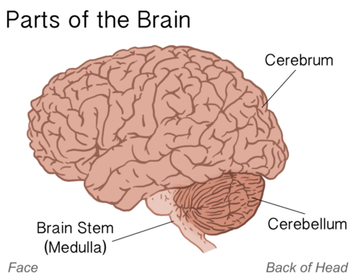
The brain controls such mental processes as reasoning, imagination, memory, and language. It also interprets information from the senses and commands the body's responses. It controls basic physiological processes such as breathing and heartbeat as well as voluntary activities such as walking and writing. The brain has three major parts: the cerebrum, cerebellum, and brainstem (Figure \(\PageIndex{3}\)). The figure shows the brain from the left side of the head. It shows how the brain would appear if the skull and meninges were removed. The brainstem links to the spinal cord via the medulla. The cerebellum is a small structure at the back of the brain. The largest part of the brain is the cerebrum.
Spinal Cord
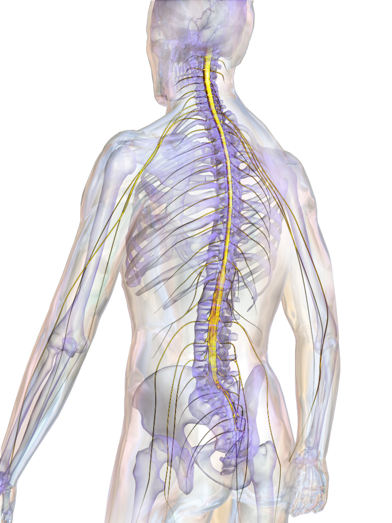
The spinal cord is a long, thin, tubular bundle of nervous tissues that extends from the brainstem down through the center of the back and pelvis. It is highlighted in yellow in Figure \(\PageIndex{15}\).
Structure of the Spinal Cord
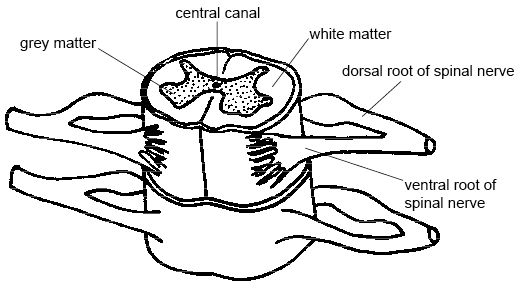
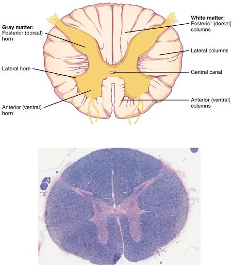
The center of the spinal cord consists of gray matter, which is made up mainly of neuron cell bodies, including interneurons and motor neurons. The gray matter is surrounded by white matter that consists mainly of myelinated axons from motor and sensory neurons. Sensory neuron axons enter the posterior side through the dorsal (/posterior) nerve root. Motor neuron axons emerge from the anterior side through the ventral (/anterior) nerve root. Note that it is common to see the terms dorsal (dorsal = back) and ventral (ventral = belly) used interchangeably with posterior and anterior, particularly in reference to nerves and the structures of the spinal cord. The central canal travels through the center of the spinal cord. The central canal is filled with cerebrospinal fluid (CSF). Figure \(\PageIndex{16}\) shows the gray matter, white matter, central canal, dorsal roots and ventral roots of the spinal cord.
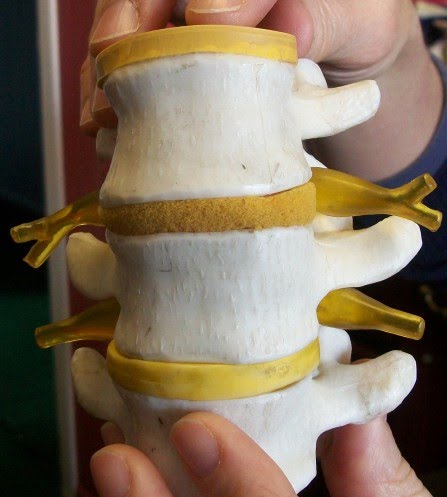
At each level of the spinal cord, the dorsal and ventral roots come together to form spinal nerves. Because the spinal cord is shorter than the vertebral column it is enclosed within, nerve roots must travel lower to exit at the associated vertebra. Spinal nerves connect the spinal cord to the PNS and enter/exit from the spinal cord between vertebrae (Figure \(\PageIndex{17}\)).
Gray Horns
In cross-section, the gray matter of the spinal cord has the appearance of an ink-blot test, with the spread of the gray matter on one side replicated on the other—a shape reminiscent of a bulbous capital “H” or a butterfly. As shown in Figure \(\PageIndex{18}\), the gray matter is subdivided into regions that are referred to as horns.
The posterior horn is responsible for sensory processing. The anterior horn sends out motor signals to the skeletal muscles. The lateral horn, which is only found in the thoracic, upper lumbar, and sacral regions, is the central component of the sympathetic division of the autonomic nervous system.
Functions of the Spinal Cord
The spinal cord serves as an information superhighway. It passes messages from the body to the brain and from the brain to the body. Sensory (afferent) nerves carry nerve impulses to the brain from sensory receptor cells everywhere in and on the body. Motor (efferent) nerves carry nerve impulses away from the brain to glands, organs, or muscles throughout the body.
The spinal cord also independently controls certain rapid responses called reflexes without any input from the brain. A sensory receptor responds to a sensation and sends a nerve impulse along a sensory nerve to the spinal cord. In the spinal cord, the message passes to an interneuron and from the interneuron to a motor nerve, which carries the impulse to a muscle. The muscle contracts in response. These neuron connections form a reflex arc, which requires no input from the brain. No doubt you have experienced such reflex actions yourself. For example, you may have reached out to touch a pot on the stove, not realizing that it was very hot. Virtually at the same moment that you feel the burning heat, you jerk your arm back and remove your hand from the pot.
Injuries to the Spinal Cord
Physical damage to the spinal cord may result in paralysis, which is a loss of sensation and movement in part of the body. Paralysis generally affects all the areas of the body below the level of the injury because nerve impulses are interrupted and can no longer travel back and forth between the brain and body beyond that point. If an injury to the spinal cord produces nothing more than swelling, the symptoms may be transient. However, if nerve fibers (axons) in the spinal cord are badly damaged, the loss of function may be permanent. Experimental studies have shown that spinal nerve fibers attempt to regrow, but tissue destruction usually produces scar tissue that cannot be penetrated by the regrowing nerves, as well as other factors that inhibit nerve fiber regrowth in the central nervous system.
Each year, many millions of people have a stroke, and stroke is the second leading cause of death in adults. Stroke, also known as cerebrovascular accident, occurs when poor blood flow to the brain results in the death of brain cells. There are two main types of strokes:
- Ischemic strokes occur due to a lack of blood flow because of a blood clot in an artery going to the brain.
- Hemorrhagic strokes occur due to bleeding from a broken blood vessel in the brain.
Either type of stroke may result in paralysis, loss of the ability to speak or comprehend speech, loss of bladder control, personality changes, and many other potential effects, depending on the part of the brain that is injured. The effects of a stroke may be mild and transient or more severe and permanent. A stroke may even be fatal. It generally depends on the type of stroke and how extensive it is.
Are you at risk of stroke? The main risk factor for stroke is age: about two-thirds of strokes occur in people over the age of 65. There is nothing you can do about your age, but most other stroke risk factors can be reduced with lifestyle changes or medications. The risk factors include high blood pressure, tobacco smoking, obesity, high blood cholesterol, diabetes mellitus, and atrial fibrillation.
Chances are good that you or someone you know is at risk of a stroke, so it is important to recognize a stroke if one occurs. Stoke is a medical emergency, and the more quickly treatment is given, the better the outcome is likely to be. In the case of ischemic strokes, the use of clot-busting drugs may prevent permanent brain damage if administered within 3 or 4 hours of the stroke. Remembering the signs of a stroke is easy. They are summed up by the acronym FAST, as explained in Figure \(\PageIndex{19}\).
Summary
The central nervous system (CNS) includes the brain and spinal cord.
The spinal cord extends from the brainstem down through the center of the back and pelvis. Sensory neuron axons enter through the dorsal (/posterior) nerve root and motor neuron axons emerge from the ventral (/anterior) nerve root. Gray matter (containing of neuron cell bodies) is located in the center of the spinal cord, surrounded by white matter (myelinated axons). The gray matter is subdivided into the posterior, anterior, and lateral horns. The spinal cord passes messages from the body to the brain and from the brain to the body. Sensory (afferent) nerves carry nerve impulses to the brain from sensory receptor cells everywhere in and on the body. Motor (efferent) nerves carry nerve impulses away from the brain to glands, organs, or muscles throughout the body. The spinal cord also independently controls reflexes without any input from the brain. Physical damage to the spinal cord may result in paralysis, which may be permanent.
References
Basinger H., & Hogg, J.P. (2021) Neuroanatomy, Brainstem. In: StatPearls [Internet]. Treasure Island (FL): StatPearls Publishing; 2022 Jan-. Available from: https://www.ncbi.nlm.nih.gov/books/NBK544297/
Lent, R., Azevedo, F.A., Andrade-Moraes, C.H., & Pinto, A.V. (2012) How many neurons do you have? Some dogmas of quantitative neuroscience under revision. European Journal of Neuroscience 35(1),1-9. doi: 10.1111/j.1460-9568.2011.07923.x. Epub 2011 Dec 13. PMID: 22151227.
Additional Resources
Watch a 3D rotating view of the sensory homunculus: Primary Somatosensory Cortes with homunculus
Watch an animated degeneration of gyri and sulci with Alzheimer's Disease: Cortical atrophy in Alzheimer's Disease
More than 40 million people worldwide suffer from Alzheimer’s disease, a brain disorder, and the number is expected to grow dramatically in the coming decades. The disease was discovered more than a century ago, but little progress has been made in finding a cure. Watch this exciting TED talk in which scientist Samuel Cohen shares a new breakthrough in Alzheimer's research as well as a message of hope that a cure for Alzheimer’s will be found.
Attributions
- Figures:
- Front of Sensory Homunculus and Rear of Sensory Homunculus by Mpj29, CC BY-SA 4.0, via Wikimedia Commons
- Brain and spinal cord: dissection, back view (coloured line engraving by W.H. Lizars, ca. 1827. Iconographic Collections) from Wellcome Images, a website operated by Wellcome Trust, a global charitable foundation based in the United Kingdom, CC BY 4.0, via Wikimedia Commons; AND Human brain and spinal cord by Z22, CC BY-SA 4.0, via Wikimedia Commons
- Spinal cord by BruceBlaus licensed CC BY 3.0 via Wikimedia Commons
- The spinal cord by Ruth Lawson Otago Polytechnic, CC BY 3.0, via Wikimedia Commons
- Spinal readjustment by Tomwsulcer dedicated CC0 via Wikimedia Commons
- Spinal cord cross section by OpenStax, CC BY 4.0, via Wikimedia Commons; Micrograph provided by the Regents of University of Michigan Medical School © 2012; CC-BY-4.0 Open Oregon, Anatomy and Physiology
- Stroke Communications Kit by CDC, public domain
- Text adapted from:
- " Central Nervous System" by Suzanne Wakim & Mandeep Grewal, LibreTexts is licensed under CC BY-NC .
- " The Cerebrum" by OpenStax, LibreTexts is licensed under CC BY.
- " The Diencephalon" by OpenStax, LibreTexts is licensed under CC BY.
- " The Brain Stem" by OpenStax, LibreTexts is licensed under CC BY.
- " The Cerebellum" by OpenStax, LibreTexts is licensed under CC BY.
- " The Spinal Cord" by OpenStax, LibreTexts is licensed under CC BY.
- " Brain- Cerebrum" by Whitney Menefee, Julie Jenks, Chiara Mazzasette, & Kim-Leiloni Nguyen, LibreTexts is licensed under CC BY .
- " Brain- Diencephalon, Brainstem, Cerebellum and Limbic System" by Whitney Menefee, Julie Jenks, Chiara Mazzasette, & Kim-Leiloni Nguyen, LibreTexts is licensed under CC BY .
- Changes: Text (and images) from above sources were pieced together with some modifications, transitions and additional content and images added by Naomi I. Gribneau Bahm, PhD., Psychology Professor at Cosumnes River College, Sacramento, CA.