
4.1: The Central Nervous System
- Page ID
- 170815
This page is a draft and under active development. Please forward any questions, comments, and/or feedback to the ASCCC OERI (oeri@asccc.org).
- Identify the three main parts of the brain
- Describe the lobes of the cerebrum and their locations
- Identify the components of the diencephalon and describe their functions
- Identify the regions of the brainstem and describe their functions
- Identify the major structures of the limbic system and describe their functions
- Describe the structure and function of the cerebellum
Overview
This module defines the central nervous system (the brain and spinal cord) and goes into detail on its components. Structures and functions of the brain are discussed, including the cerebrum: hemispheres and lateralization, the cerebral cortex, the frontal, parietal, temporal and occipital lobes; the brainstem: diencephalon, midbrain, pons and medulla; the limbic system; and the cerebellum; as well as structures, function and injury of the spinal cord.
Figure \(\PageIndex{1}\) is a very odd-looking model called a homunculus. This model shows the relative representation of different parts of the body in the primary sensory cortex, an area in the parietal lobe of the brain. As you can see, larger areas of the brain in this region are associated with the hands, face, and tongue than the arms, torso, legs and feet. Larger areas of representation in the brain result in greater sensitivity in those body areas. Given the importance of speech, manual dexterity, and face-to-face social interactions in human beings, it is not surprising that relatively large areas of the brain are needed to relay sensation from these body parts. (There is a similarly odd-looking representation of voluntary motor functions called the motor homunculus that is located in the primary motor cortex, an area in the frontal lobe of the brain.) The brain is the most complex organ in the human body and is part of the central nervous system.
Cerebrum
The iconic gray mantle of the human brain, which appears to make up most of the mass of the brain, is the cerebrum. It controls conscious, intellectual functions, such as reasoning, language, memory, sight, touch, and hearing. When you read a book, play a video game, or recognize a classmate, you are using your cerebrum. The wrinkled portion is the cerebral cortex (see below), and the rest of the structure is deep to that outer covering.
Hemispheres and Lateralization of the Cerebrum
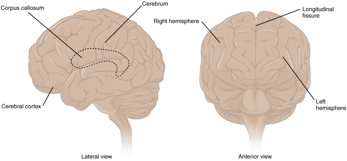
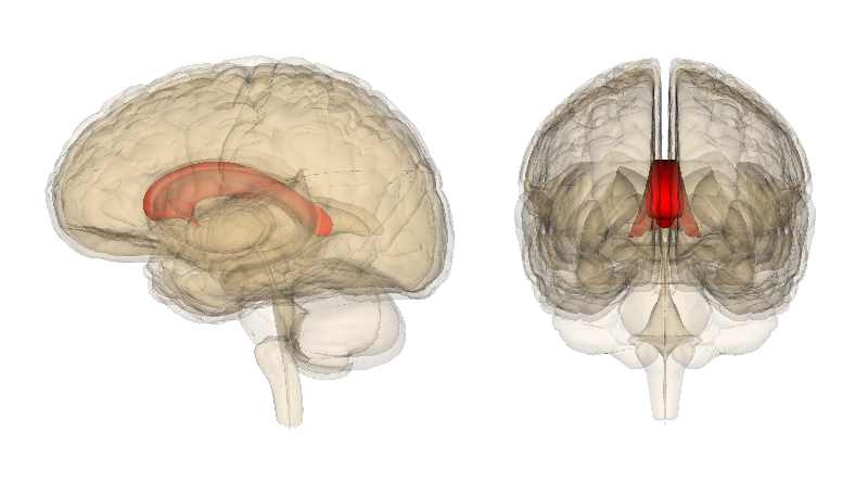
There is a large separation between the two sides of the cerebrum called the longitudinal fissure. It separates the cerebrum into two distinct halves, the right and left cerebral hemispheres. The two hemispheres are connected by a thick bundle of axons, known as the corpus callosum, which lies deep within the cerebrum. The corpus callosum is the major pathway for communication between the two hemispheres, connecting each point in the cerebral cortex to the mirror-image point in the opposite hemisphere. The location of the corpus callosum is shown with a dotted outline in Figure \(\PageIndex{4}\), as though you are looking through the outside and deep inside the brain. Figure \(\PageIndex{5}\) shows a three-dimensional image of the corpus callosum in relation to other deep brain structures and surrounded by the cerebrum.
The right and left hemispheres of the cerebrum are similar in shape, and most areas of the cerebrum are found in both hemispheres. Some areas, however, show lateralization, or a concentration in one hemisphere or the other. For example, in most people, language functions are more concentrated in the left hemisphere, whereas abstract reasoning and visual-spatial abilities are more concentrated in the right hemisphere.
For reasons that are not yet clear, each hemisphere of the brain interacts primarily with the contralateral (opposite) side of the body. The left side of the brain receives messages from and sends commands to the right side of the body, and the right side of the brain receives messages from and sends commands to the left side of the body. Sensory nerves from the spinal cord to the brain and motor nerves from the brain to the spinal cord both cross to the other side of the body.
Cerebral Cortex
The cerebrum is covered by a continuous layer of gray matter that wraps around either side of the forebrain—the cerebral cortex. This thin (just a few millimeters thick), extensive region of wrinkled gray matter is responsible for most of the information processing and higher functions of the nervous system. The cerebral cortex has many folds in it that greatly increase the surface area of the brain. A gyrus (plural = gyri) is the ridge of one of those wrinkles, and a sulcus (plural = sulci) is the groove between two gyri. The pattern of these folds of tissue indicates specific regions of the cerebral cortex. At birth, the head of the newborn is limited by the size of the birth canal, and the brain must fit inside the cranial cavity of the skull. Extensive folding in the cerebral cortex enables more gray matter to fit into this limited space. If the gray matter of the cerebral cortex in an adult were peeled off of the cerebrum and laid out flat, its surface area would be about 2,500 cm2 (2.5 ft2), or about the size of the top of a card table.
Lobes of the Cerebral Cortex
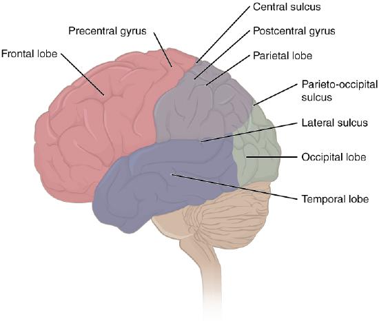
The surface of the brain can be mapped on the basis of the locations of large gyri and sulci. Using these landmarks, the cortex can be separated into four major regions, or lobes (Figure \(\PageIndex{6}\)). The lateral sulcus that separates the temporal lobe from the other regions is one such landmark.
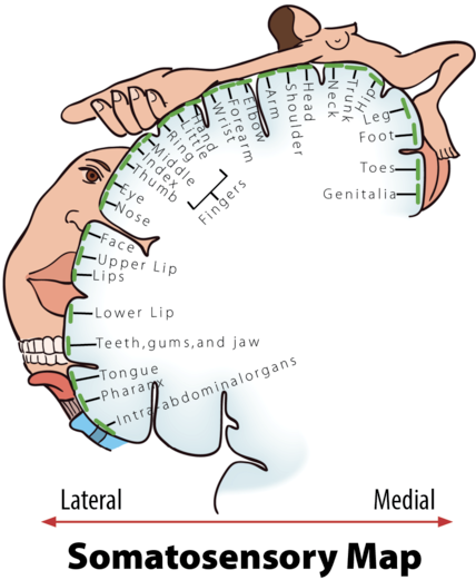
Superior to the lateral sulcus are the frontal lobe and parietal lobe, which are separated from each other by the central sulcus. The most posterior gyrus of the frontal lobe is the precentral gyrus, which is where the primary motor cortex is located, while the most anterior gyrus of the parietal lobe is the postcentral gyrus, which is where the primary somatosensory cortex is located. (The primary somatosensory cortex is the area that the homonculus in Figure \(\PageIndex{1}\) represents.)
The posterior region of the cortex is the occipital lobe, which has no obvious anatomical border between it and the parietal or temporal lobes on the lateral surface of the brain. From the medial surface, the parieto-occipital sulcus separates the parietal and occipital lobes.
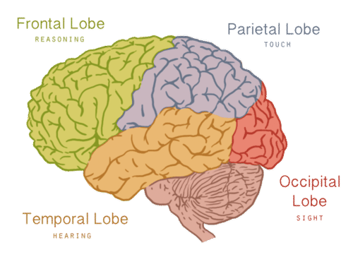
Figure \(\PageIndex{7}\) has only the four lobes labeled for quicker reference. The lobes are associated with multiple functions. The image shows one function of each lobe: frontal is associated with reasoning, parietal with touch, occipital with sight, and temporal with hearing. The lobes (found on each hemisphere of the cerebrum) are described in greater detail below.
Frontal Lobe
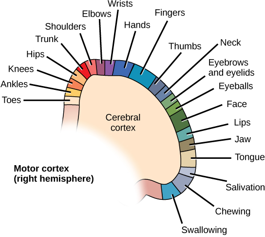
The frontal lobes are located at the front of the brain behind the forehead. The frontal lobes are associated with executive functions such as attention, self-control, planning, problem-solving, reasoning, abstract thought, language, and personality. Another important function of the frontal lobes is movement- as mentioned above, the precentral gyrus (indicated on Figure \(\PageIndex{6}\)) contains the primary motor cortex. Like the sensory homunculus pictured in the beginning of this section (Figure \(\PageIndex{1}\)), there is a similarly disproportionate representation of voluntary motor control (the motor homunculus) located in the precentral gyrus. Figure \(\PageIndex{8}\) shows the areas of the primary motor cortex dedicated to movement of various body areas. Notice that like the sensory homunculus, the hands, fingers, thumb, and facial areas have a greater share of the cortex than areas with less fine-tuned movement (like the shoulders, trunk, and hips, for example).
Parietal Lobe
The parietal lobes are located posterior to the frontal lobes at the top of the head. The parietal lobes are involved in body sensations, including temperature, touch, and pain. As mentioned above, the postcentral gyrus (indicated on Figure \(\PageIndex{6}\)) contains the primary somatosensory cortex. The sensory homunculus model pictured in the beginning of this section (Figure \(\PageIndex{1}\)) shows the strange character that results from making the body areas proportional to their representation in the brain. The drawing in Figure \(\PageIndex{9}\) shows the sensory homunculus stretched out along the postcentral gyrus of the parietal lobe. Notice that areas that have sensation but no voluntary motor control (like the genitals and the intra-abdominal organs) are present on the primary somatosensory cortex but not on the primary motor cortex (Figure \(\PageIndex{8}\)). Reading and arithmetic are also functions of the parietal lobes.
Temporal Lobe
The temporal lobes are located at the sides of the head below the frontal and parietal lobes. The temporal lobes enable the formation and retrieval of memories, the integration of memories and sensations, and perception of the senses of hearing (audition), and smell (olfaction)- primary auditory cortex and primary olfactory cortex are both located in the temporal lobes.
Occipital Lobe
The occipital lobes are located at the back of the head below the parietal lobes. The occipital lobes are the smallest of the four pairs of lobes. Primary visual cortex is located in the occipital lobes, and they are dedicated almost solely to vision.
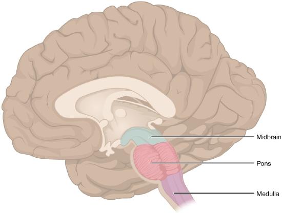
Brainstem
As noted separately (see the nervous system structure module), sometimes the diencephalon is considered as a separate additional region, and sometimes it is included as the uppermost part of the brainstem (Basinger and Hogg, 2021), as it is here. The brainstem is the central region of the brain that connects the cerebrum (on the superior end) to the diencephalon, midbrain, pons, medulla, and ultimately joins the spinal cord (on the inferior end). The cerebellum is attached to the brainstem, but is considered a separate region of the adult brain.
One of the brainstem’s most important roles is that of an “information highway.” That is, all of the information coming from the body to the brain and the information from the cerebrum to the body go through the brainstem. Sensory pathways for such things as pain, temperature, touch, and pressure sensation go upward to the cerebrum, and motor pathways for movement and other body processes go downward to the spinal cord. Most of the axons in the motor pathways cross from one side of the CNS to the other as they pass through the medulla. As a result, the right side of the brain controls much of the movement on the left side of the body, and the left side of the brain controls much of the movement on the right side of the body. Similarly, axons in sensory pathways cross from one side of the CNS to the other either in the spinal cord or in the medulla (depending on the specific sensory information carried, such as pain versus localized touch), such that the right side of the brain receives sensation primarily from the left side of the body and vice versa.
Diencephalon
The diencephalon ("through brain") is the one region of the adult brain that retains its name from embryonic development. It connects the cerebrum and the rest of the nervous system, with one exception. The brainstem, the spinal cord, and the PNS all send information to the cerebrum through the diencephalon, and output from the cerebrum then passes back through the diencephalon. The single exception to this pattern is the sense of smell (olfaction), which connects directly to the cerebrum first. The diencephalon comprises the ventral surface of the forebrain (and connects to the tapering cone of the midbrain, pons, and medulla, the remaining structures in the brainstem).
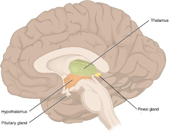
The diencephalon is located deep within the cerebrum and constitutes the walls of the third ventricle. The diencephalon can be described as any region of the brain with “thalamus” in its name. The two major regions of the diencephalon are the thalamus itself and the hypothalamus (Figure \(\PageIndex{10}\)). There are other structures as well, such as the epithalamus, which contains the pineal gland.
Thalamus
The thalamus, which is located above the hypothalamus (Figure \(\PageIndex{10}\) and Figure \(\PageIndex{11}\)), is a major hub for information traveling back and forth between the spinal cord, brainstem, and cerebrum. It filters sensory information traveling to the cerebrum. It relays sensory signals to the cerebral cortex and motor signals to the spinal cord. It is also involved in the regulation of consciousness, sleep, and alertness. As noted above, olfaction (smell) is the only sense that travels directly to the cortex and does not travel through the thalamus (as part of the diencephalon) first. However, olfactory messages do travel from the cortex back to the thalamus, so smell is still a participant in the "sensory relay" functions of the thalamus.
Hypothalamus
The hypothalamus ("below thalamus") is about the size of an almond and is responsible for certain metabolic processes and other activities of the autonomic nervous system, including body temperature, heart rate, hunger, thirst, fatigue, sleep, wakefulness, and circadian (24-hour) rhythms. The hypothalamus is also an important emotional center of the brain. The hypothalamus can regulate so many body functions because it responds to many different internal and external signals, including messages from the brain, light, steroid hormones, stress, and invading pathogens, among others.
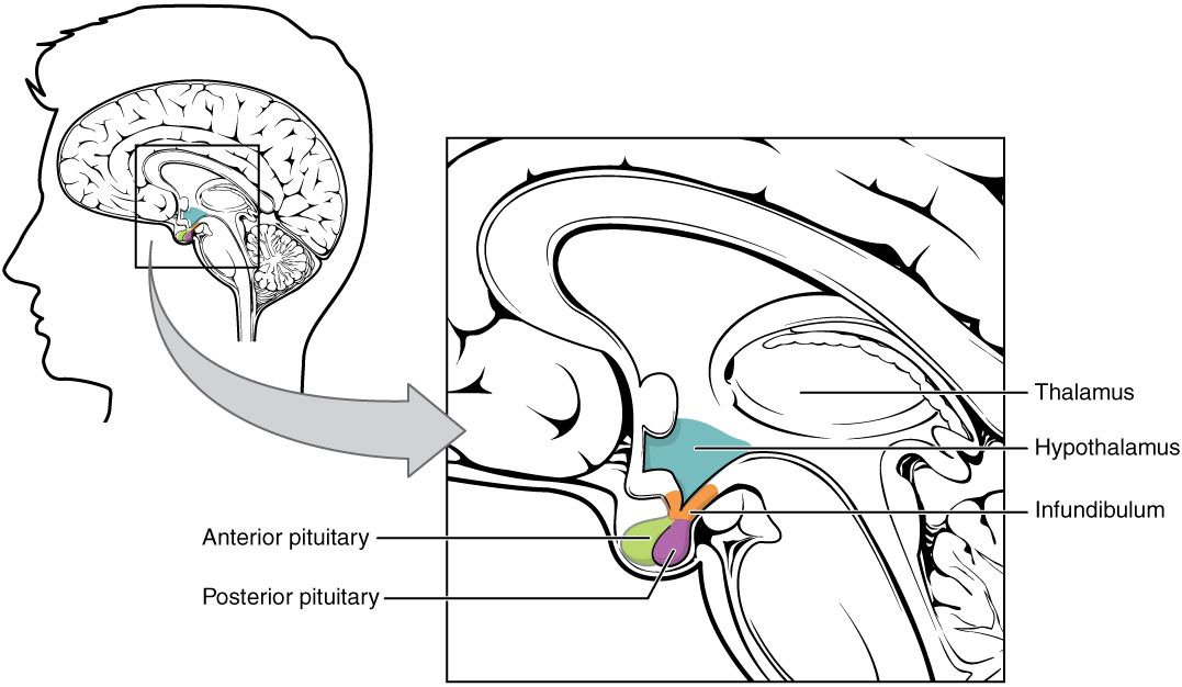
One way the hypothalamus influences body functions is by synthesizing hormones that directly influence body processes. For example, it synthesizes the hormone oxytocin, which stimulates penile and vaginal contractions during orgasm, uterine contractions during childbirth, and the letdown of milk during lactation. It also synthesizes the hormone vasopressin (also called antidiuretic hormone), which stimulates the kidneys to reabsorb more water and excrete more concentrated urine. These two hormones are sent from the hypothalamus via a stalk-like structure called the infundibulum ("pituitary stalk", see Figure \(\PageIndex{11}\)) directly to the posterior (back) portion of the pituitary gland, which secretes them into the blood.
The main way the hypothalamus influences body functions is by controlling the pituitary gland, known as the "master gland" of the endocrine system. (The endocrine system is considered separately in the communication and the endocrine system module.) The hypothalamus synthesizes neurohormones called releasing factors that travel through the infundibulum directly to the anterior (front) part of the pituitary gland. The releasing factors generally either stimulate or inhibit the secretion of anterior pituitary hormones, most of which control other glands of the endocrine system.
Figure \(\PageIndex{11}\) shows where the thalamus, hypothalamus, and pituitary gland (typically considered an extension of the hypothalamus) are located in the brain. The cerebrum, hypothalamus, and thalamus exist in two halves, one in each hemisphere. The pituitary gland is a single structure in the center, connected to the hypothalamus by the infundibulum (or "pituitary stalk").
The remaining parts to the brainstem are the midbrain, the pons, and the medulla, which are shown in Figure \(\PageIndex{12}\) below. These structures are primarily involved in the unconscious functions of the autonomic nervous system as well as several types of sensory information. They also help coordinate large body movements such as walking and running.
Midbrain
The midbrain coordinates sensory representations of sight (visual system- the superior colliculi), sound (auditory system- the inferior colliculi), and somatosensory perceptual spaces, translating these inputs before sending them to the forebrain.
Pons
The pons ("bridge") is the main connection with the cerebellum. It relays messages between other parts of the brain (primarily the cerebrum and cerebellum) and sends messages to paralyze major body muscles during REM sleep. Some researchers have hypothesized that the pons plays a role in dreaming. Some of the functions of the pons are shared by the medulla, such as regulating several crucial functions including cardiovascular system and respiratory systems, and heartbeat and breathing rates.
Medulla
The medulla (also called the medulla oblongata) controls several subconscious homeostatic functions such as breathing, heart and blood vessel activity, swallowing, and digestion.
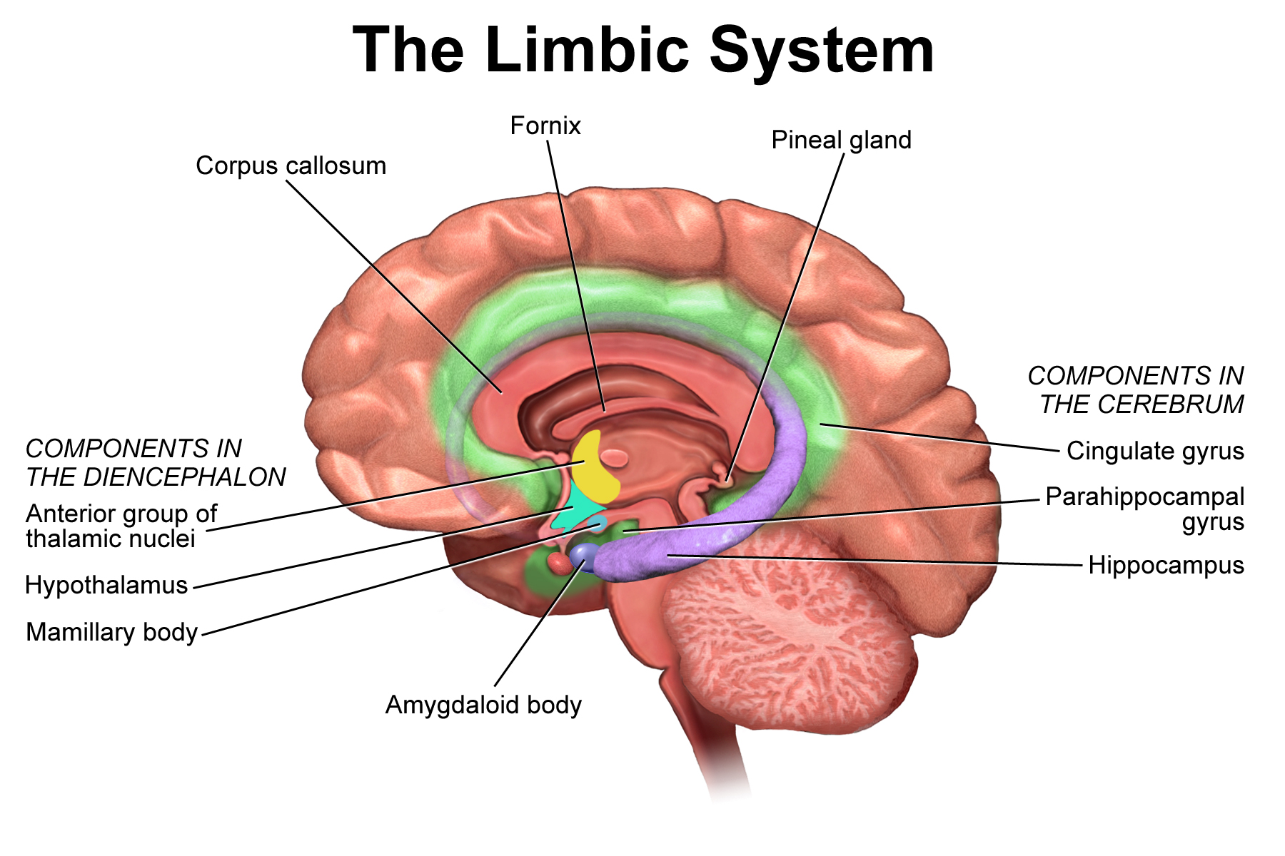
Limbic System
The limbic system is a collection of structures of the cerebrum and diencephalon that are involved in emotion, motivation and memory. Although still under debate, the structures most often recognized as belonging to this system are the cingulate gyrus, hippocampus, amygdala, olfactory structures, and various nuclei of the diencephalon. Here we will focus on the cingulate gyrus, the hippocampus, and the amygdala (Figure \(\PageIndex{13}\)). The cingulate gyrus (colored green in the figure) is located on the midsagittal surface of the cortex, immediately superior to the corpus callosum and surrounding the diencephalon. The cingulate gyrus focuses attention on events that are emotionally salient. The hippocampus (colored purple in the figure) is a nucleus shaped like a seahorse, hence its name. The hippocampus is essential for forming memories and storing them long-term and is located deep in the temporal lobe. Lastly, the amygdala (also called the amygdaloid body, colored blue in the figure) is connected to the anterior end of the hippocampus and is involved in multiple aspects of emotion, including encoding memories related to highly emotional states. The amygdala is especially important for the emotion fear.
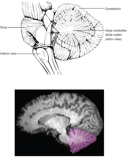
Cerebellum
The cerebellum, as the name suggests, is the “little brain”, just below the cerebrum and at the back of the brain behind the brainstem. It is covered in gyri and sulci like the cerebrum, and looks like a miniature version of that part of the brain Figure \(\PageIndex{14}\). The cerebellum is largely responsible for comparing information from the cerebrum with sensory feedback from the periphery through the spinal cord. It accounts for approximately 10 percent of the mass of the brain. The cerebellum coordinates body movements and is involved in movements that are learned with repeated practice. For example, when you hit a softball with a bat or touch type on a keyboard you are using your cerebellum. Many nerve pathways link the cerebellum with motor neurons throughout the body.
Summary
The central nervous system (CNS) includes the brain and spinal cord, described in greater detail in this module. The adult brain is separated into three major regions: the cerebrum, the brainstem, and the cerebellum. The cerebrum is the largest portion and is divided into two halves called hemispheres by the longitudinal fissure. The two hemispheres are connected by the corpus callosum, the major pathway for communication between the hemispheres.
The cerebral cortex, a continuous layer of gray matter covering the cerebrum, is the location of important cognitive functions. The cerebral cortex is separated into the frontal, parietal, temporal, and occipital lobes. The frontal lobe is responsible for executive functions and movements. The parietal lobe is involved in body sensations, reading, and arithmetic. The temporal lobe has regions crucial for memory formation, and contains cortical areas that process audition (hearing) and olfaction (smell). The occipital lobe is where visual processing begins.
The brainstem is composed of the diencephalon, midbrain, pons, and medulla. The brainstem serves as an "information highway", since all of the information coming from the body to the brain and the information from the cerebrum to the body go through the brainstem. The two major regions of the diencephalon are the thalamus and the hypothalamus. The thalamus is a relay between the cerebrum and the rest of the nervous system. The hypothalamus coordinates metabolic processes through the autonomic and endocrine systems. The midbrain coordinates sensory representations of sight, sound and somatosensation. The pons is the main connection between the cerebrum and the cerebellum. The pons and the medulla together regulate the cardiovascular and respiratory systems. The medulla also has important functions for swallowing and digestion.
The limbic system includes structures of the cerebrum and diencephalon that are responsible for emotion, motivation, and memory. The main structures are the cingulate gyrus, the hippocampus and the amygdala.
The cerebellum is connected to the brain stem and compares information from the cerebrum with sensory feedback from the spinal cord. The cerebellum coordinates body movements and is involved in movements that are learned with repeated practice.
References
Basinger H., & Hogg, J.P. (2021) Neuroanatomy, Brainstem. In: StatPearls [Internet]. Treasure Island (FL): StatPearls Publishing; 2022 Jan-. Available from: https://www.ncbi.nlm.nih.gov/books/NBK544297/
Lent, R., Azevedo, F.A., Andrade-Moraes, C.H., & Pinto, A.V. (2012) How many neurons do you have? Some dogmas of quantitative neuroscience under revision. European Journal of Neuroscience 35(1),1-9. doi: 10.1111/j.1460-9568.2011.07923.x. Epub 2011 Dec 13. PMID: 22151227.
Attributions
- Figures:
- Brain by Laura Guerin, CC BY-NC 3.0 via CK-12
- Cerebrum by OpenStax, CC BY 4.0, via Wikimedia Commons
- Corpus callosum by Images generated by Life Science Databases (LSDB), CC BY-SA 2.1 JP, via Wikimedia Commons
- Lobes of Cerebral Cortex by OpenStax, CC BY 4.0, via Wikimedia Commons
- Brain lobes by Laura Guerin, CC BY-NC 3.0 via CK-12
- Primary motor cortex by CNX OpenStax, CC BY 4.0, via Wikimedia Commons
- Somatosensory Map from NOBA module Human Sexual Anatomy and Physiology by Lucas, D. & Fox, J., CC BY-NC-SA 4.0, via NOBA
- Thalamus/hypothalamus adapted by Naomi Bahm (areas adjusted, pineal gland added) from Diencephalon by OpenStax, CC BY 4.0, via Wikimedia Commons
- Hypothalamus-Pituitary Complex by OpenStax, licensed CC BY 3.0 via Wikimedia Commons
- Brainstem adapted by Naomi Bahm (midbrain extended to include colliculi) from Brain stem by OpenStax, licensed CC BY 4.0 via Wikimedia Commons
- Limbic system by BruceBlaus licensed CC BY 3.0 via Wikimedia Commons
- Cerebellum by OpenStax, CC BY 4.0, via Wikimedia Commons; Note: MRI originally by Semiconscious, Public domain, via Wikimedia Commons
- Text adapted from:
- " Central Nervous System" by Suzanne Wakim & Mandeep Grewal, LibreTexts is licensed under CC BY-NC .
- " The Cerebrum" by OpenStax, LibreTexts is licensed under CC BY.
- " The Diencephalon" by OpenStax, LibreTexts is licensed under CC BY.
- " The Brain Stem" by OpenStax, LibreTexts is licensed under CC BY.
- " The Cerebellum" by OpenStax, LibreTexts is licensed under CC BY.
- " The Spinal Cord" by OpenStax, LibreTexts is licensed under CC BY.
- " Brain- Cerebrum" by Whitney Menefee, Julie Jenks, Chiara Mazzasette, & Kim-Leiloni Nguyen, LibreTexts is licensed under CC BY .
- " Brain- Diencephalon, Brainstem, Cerebellum and Limbic System" by Whitney Menefee, Julie Jenks, Chiara Mazzasette, & Kim-Leiloni Nguyen, LibreTexts is licensed under CC BY .
- Changes: Text (and images) from above sources were pieced together with some modifications, transitions and additional content and images added by Naomi I. Gribneau Bahm, PhD., Psychology Professor at Cosumnes River College, Sacramento, CA.